正在加载图片...
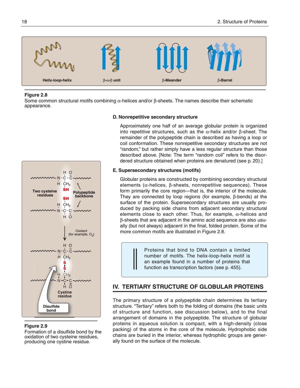
18 2.Structure of Proteins M) N 修计i Helix-oop-helix B-g-B unit B-Meander B-Barrel Figure 2.8 structural motifs mbining o-helices and/or B-sheets.The names describe their schematic D.Nonrepetitive secondary structure Approximately one half of an average globular protein is organized mpetive structures,such as the a-helix and/or B-she as having a loop o random.but rather simply have a less regular structure than those described above.[Note:The term"random coil"refers to the disor dered structure obtained when proteins are denatured (see p.20).] E.Supersecondary structures(motifs) Globular proteins are constructed by combining secondary structural elements (a-helices,B-sheets,nonrepetitive sequences).These TwoSXtene are connected by oop( the CH duced by packing side chains from adjacent secondary structural B-shes to oach other.Thus,for example.a-helices and 0ac more common motifsare iustrated in Fiqure 28 wN-c AAAV Proteins that bind to numt function as transcription factors(see p.455). IV.TERTIARY STRUCTURE OF GLOBULAR PROTEINS The primary structure of a polypeptide chain determines its tertiary e folding of domains (the basic units arr of de id n The str Figure 2.9 proteninquouiniscomwithaigdensity(cls producing one cystine residue D. Nonrepetitive secondary structure Approximately one half of an average globular protein is organized into repetitive structures, such as the α-helix and/or β-sheet. The remainder of the polypeptide chain is described as having a loop or coil conformation. These nonrepetitive secondary structures are not “random,” but rather simply have a less regular structure than those described above. [Note: The term “random coil” refers to the disordered structure obtained when proteins are denatured (see p. 20).] E. Supersecondary structures (motifs) Globular proteins are constructed by combining secondary structural elements (α-helices, β-sheets, nonrepetitive sequences). These form primarily the core region—that is, the interior of the molecule. They are connected by loop regions (for example, β-bends) at the surface of the protein. Supersecondary structures are usually produced by packing side chains from adjacent secondary structural elements close to each other. Thus, for example, α-helices and β-sheets that are adjacent in the amino acid sequence are also usually (but not always) adjacent in the final, folded protein. Some of the more common motifs are illustrated in Figure 2.8. Proteins that bind to DNA contain a limited number of motifs. The helix-loop-helix motif is an example found in a number of proteins that function as transcription factors (see p. 455). IV. TERTIARY STRUCTURE OF GLOBULAR PROTEINS The primary structure of a polypeptide chain determines its tertiary structure. “Tertiary” refers both to the folding of domains (the basic units of structure and function, see discussion below), and to the final arrangement of domains in the polypeptide. The structure of globular proteins in aqueous solution is compact, with a high-density (close packing) of the atoms in the core of the molecule. Hydrophobic side chains are buried in the interior, whereas hydro philic groups are generally found on the surface of the molecule. Figure 2.8 Some common structural motifs combining α-helices and/or β-sheets. The names describe their schematic appearance. Helix-loop-helix β-α-β unit β-Meander β-Barrel 18 2. Structure of Proteins Figure 2.9 Formation of a disulfide bond by the oxidation of two cysteine residues, producing one cystine residue. C C H CH2 N O H Two cysteine residues H N C H CH2 SH SH C C H C O CH2 N O H Polypeptide backbone Disulfide bond Oxidant (for example, O2) 168397_P013-024.qxd7.0:02 Protein structure 5-20-04 2010.4.4 11:31 AM Page 18