正在加载图片...

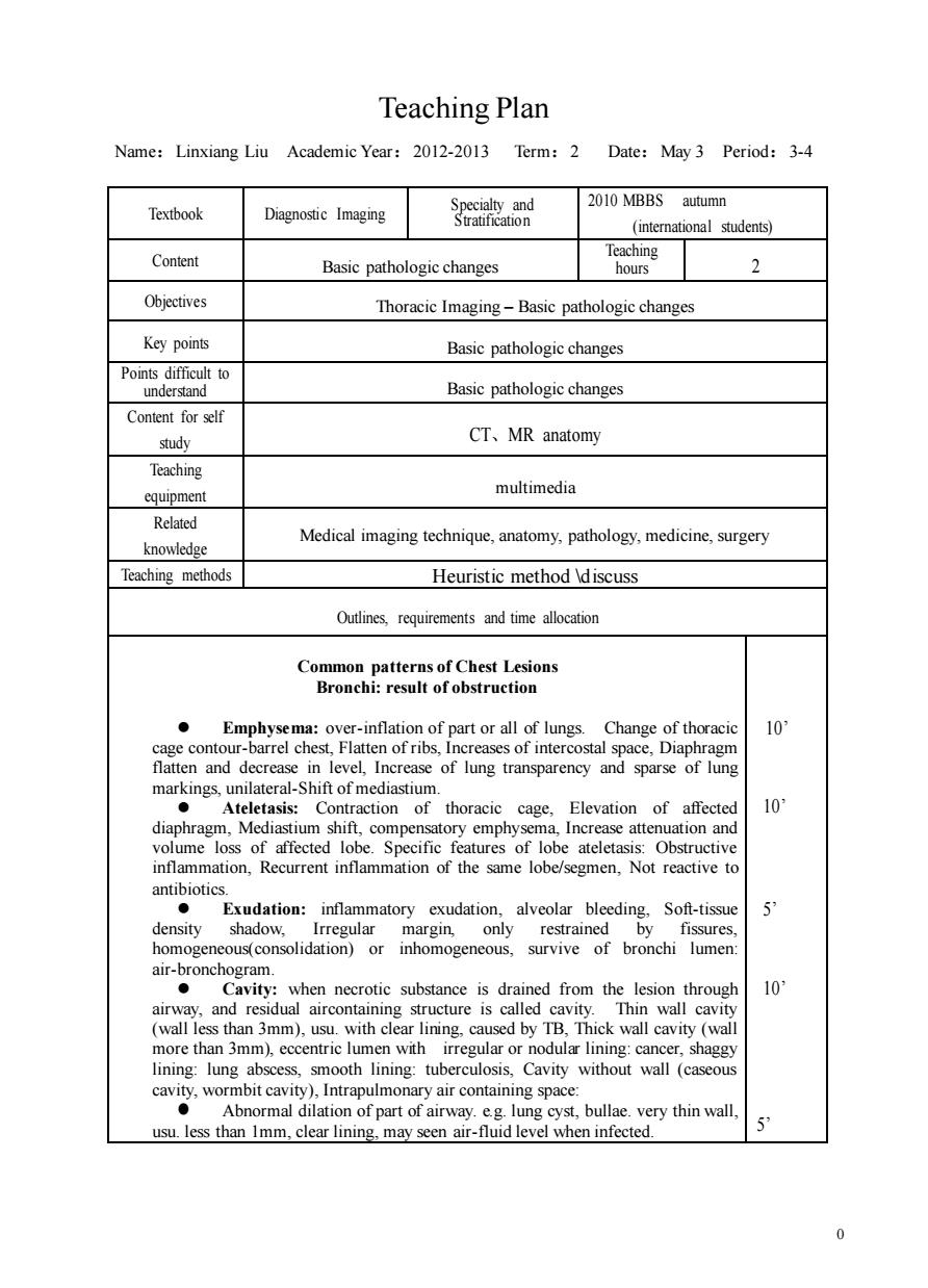
Teaching Plan Name:Linxiang Liu Academic Year:2012-2013 Term:2 Date:May3 Period:3-4 2010 MBBS autumn Textbook Diagnostic Imaging Suaion (students) Content Basic pathologic changes Teaching 2 Objectives Thoracic Imaging-Basic pathologic changes Key points Basic pathologic changes Points difficult to under Basic pathologic changes Content for self study CT、MR anatomy Teaching equipment multimedia Related knowledge Medical imaging technique,anatomy,pathology,medicine,surgery Teaching methods Heuristic method discuss Outlines,requirements and time allocation Common patterns of Chest Lesions Bronchi:result of obstruction ver-inlationofpartoralofluneg Cha 10 ostal sn flatten and decrease in level,Increase of lung transparency and sparse of lung markings,unilateral-Shift of mediastium. Contraction of thoracic cage, Elevation of affected 10 g of affo inflammation,Recurrent inflammation of the same lobe/segmen,Not reactive to antibiotics. Exudation:inflammatory exudation,alveolar bleeding,Soft-tissue 5 density shadow, by ty:when necrotic substance is drained from the lesion through 10 airway,and residual aircontaining structure is called cavity.Thin wall cavity (wall less than 3mm),usu.with clear lining.caused by TB,Thick wall cavity (wall more th an 3mm),eccentric lumen with irregular or lining:lung abs ooth lining:tut yair cont usu.less than Imm.clear linin bullae.very thin wall. enar-fuid level when infected 0 0 Teaching Plan Name:Linxiang Liu Academic Year:2012-2013 Term:2 Date:May 3 Period:3-4 Textbook Diagnostic Imaging Specialty and Stratification 2010 MBBS autumn (international students) Content Basic pathologic changes Teaching hours 2 Objectives Thoracic Imaging – Basic pathologic changes Key points Basic pathologic changes Points difficult to understand Basic pathologic changes Content for self study CT、MR anatomy Teaching equipment multimedia Related knowledge Medical imaging technique, anatomy, pathology, medicine, surgery Teaching methods Heuristic method \discuss Outlines, requirements and time allocation Common patterns of Chest Lesions Bronchi: result of obstruction ⚫ Emphysema: over-inflation of part or all of lungs. Change of thoracic cage contour-barrel chest, Flatten of ribs, Increases of intercostal space, Diaphragm flatten and decrease in level, Increase of lung transparency and sparse of lung markings, unilateral-Shift of mediastium. ⚫ Ateletasis: Contraction of thoracic cage, Elevation of affected diaphragm, Mediastium shift, compensatory emphysema, Increase attenuation and volume loss of affected lobe. Specific features of lobe ateletasis: Obstructive inflammation, Recurrent inflammation of the same lobe/segmen, Not reactive to antibiotics. ⚫ Exudation: inflammatory exudation, alveolar bleeding, Soft-tissue density shadow, Irregular margin, only restrained by fissures, homogeneous(consolidation) or inhomogeneous, survive of bronchi lumen: air-bronchogram. ⚫ Cavity: when necrotic substance is drained from the lesion through airway, and residual aircontaining structure is called cavity. Thin wall cavity (wall less than 3mm), usu. with clear lining, caused by TB, Thick wall cavity (wall more than 3mm), eccentric lumen with irregular or nodular lining: cancer, shaggy lining: lung abscess, smooth lining: tuberculosis, Cavity without wall (caseous cavity, wormbit cavity), Intrapulmonary air containing space: ⚫ Abnormal dilation of part of airway. e.g. lung cyst, bullae. very thin wall, usu. less than 1mm, clear lining, may seen air-fluid level when infected. 10’ 10’ 5’ 10’ 5’