正在加载图片...
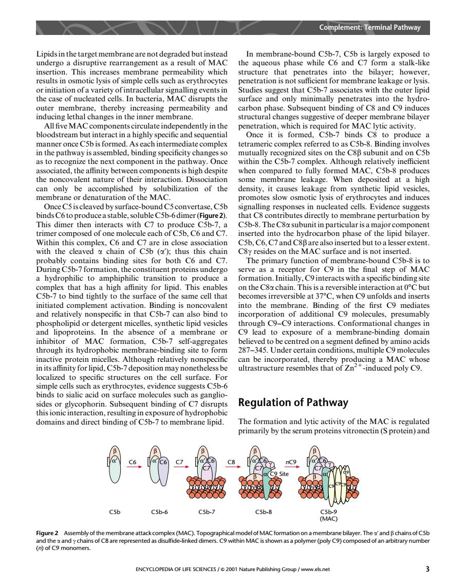
Complement:Terminal Pathway Lipidsin the target membrane are not degraded but instead In membrane-bound C5b-7.C5b is largely exposed to undergo a disruptive rearrangement as a result of MAC the aqueous phase while C6 and C7 form a stalk-like insertion. This s men tructure that penetrates into the bilayer:however. hat C5h for memt the the case of nucleated cells.In bacteria.MAC disrupts the surface and only minimally penetrates into the hydro outer membrane,thereby increasing permeability and carbon phase.Subsequent binding of C8 and C9 induces I changes in the inner bilayer L late in it is for manner once C5b is formed.As each intermediate complex tetrameric complex referred to as C5b-8.Binding involves in the pathway is assembled,binding specificity changesso and on C5b as to o ext component in the pa ay. the n t alent nat der osited at a can only be accomplished by solubilization of the density.it causes leakage from synthetic lipid vesicles membrane or denaturation of the MAC promotes slow osmotic lysis of erythrocytes and induces Oncecroproducsastable solubiecsh sim se,C5 ponses n n eated cells. ith C C5b-8.The ctly to m inserted into the hydrocarbon phase of the lipid bilayer rextent the on th urface and is no as mary C9h the final of MAC a hydrophilic to amphiphilic transition to produce e on the C8chain. th and relatively nonspecific in that Csh- bind to onal C9 phospholipid or detergent micelles,synthetic lipid vesicles gh C9-C9interactions.Conformational changes in and lipoproteins. In the absence of a membrane lead to exposure of a membrane-binding domain inhibit MA self-aggregate centred on a egment den be ted ordCs-7depitb MAC wh localized to spec cifc structures on the cell surface. as erythrocytes, idence sugges ent binding of C7 disr Regulation of Pathway thisionic interaction,resulting in exposure of hydrophobic domains and direct binding of C5b-7 to membrane lipid. The formation and lytic activity of the MAC is regulatec primarly by the serum proteins vitronectin (S protein)and nC9 C5b-6 C5b-7 C5b-8 Figure2 Assembly of th yer.Theand B chains ofCsb s of C8 are represented as of an arbitrary numb ENCYCLOPEDIA OF LIFE SCIENCES/e2001 Natu Publishing Group /www.els.neLipids in the target membrane are not degraded but instead undergo a disruptive rearrangement as a result of MAC insertion. This increases membrane permeability which results in osmotic lysis of simple cells such as erythrocytes or initiation of a variety of intracellular signalling events in the case of nucleated cells. In bacteria, MACdisrupts the outer membrane, thereby increasing permeability and inducing lethal changes in the inner membrane. All five MACcomponents circulate independently in the bloodstream but interact in a highly specific and sequential manner once C5b is formed. As each intermediate complex in the pathway is assembled, binding specificity changes so as to recognize the next component in the pathway. Once associated, the affinity between components is high despite the noncovalent nature of their interaction. Dissociation can only be accomplished by solubilization of the membrane or denaturation of the MAC. Once C5 is cleaved by surface-bound C5 convertase, C5b binds C6 to produce a stable, soluble C5b-6 dimer (Figure2). This dimer then interacts with C7 to produce C5b-7, a trimer composed of one molecule each of C5b, C6 and C7. Within this complex, C6 and C7 are in close association with the cleaved a chain of C5b (a’); thus this chain probably contains binding sites for both C6 and C7. During C5b-7 formation, the constituent proteins undergo a hydrophilic to amphiphilic transition to produce a complex that has a high affinity for lipid. This enables C5b-7 to bind tightly to the surface of the same cell that initiated complement activation. Binding is noncovalent and relatively nonspecific in that C5b-7 can also bind to phospholipid or detergent micelles, synthetic lipid vesicles and lipoproteins. In the absence of a membrane or inhibitor of MACformation, C5b-7 self-aggregates through its hydrophobic membrane-binding site to form inactive protein micelles. Although relatively nonspecific in its affinity for lipid, C5b-7 deposition may nonetheless be localized to specific structures on the cell surface. For simple cells such as erythrocytes, evidence suggests C5b-6 binds to sialic acid on surface molecules such as gangliosides or glycophorin. Subsequent binding of C7 disrupts this ionic interaction, resulting in exposure of hydrophobic domains and direct binding of C5b-7 to membrane lipid. In membrane-bound C5b-7, C5b is largely exposed to the aqueous phase while C6 and C7 form a stalk-like structure that penetrates into the bilayer; however, penetration is not sufficient for membrane leakage or lysis. Studies suggest that C5b-7 associates with the outer lipid surface and only minimally penetrates into the hydrocarbon phase. Subsequent binding of C8 and C9 induces structural changes suggestive of deeper membrane bilayer penetration, which is required for MAClytic activity. Once it is formed, C5b-7 binds C8 to produce a tetrameric complex referred to as C5b-8. Binding involves mutually recognized sites on the C8b subunit and on C5b within the C5b-7 complex. Although relatively inefficient when compared to fully formed MAC, C5b-8 produces some membrane leakage. When deposited at a high density, it causes leakage from synthetic lipid vesicles, promotes slow osmotic lysis of erythrocytes and induces signalling responses in nucleated cells. Evidence suggests that C8 contributes directly to membrane perturbation by C5b-8. The C8a subunit in particular is a major component inserted into the hydrocarbon phase of the lipid bilayer. C5b, C6, C7 and C8b are also inserted but to a lesser extent. C8g resides on the MACsurface and is not inserted. The primary function of membrane-bound C5b-8 is to serve as a receptor for C9 in the final step of MAC formation. Initially, C9 interacts with a specific binding site on the C8a chain. This is a reversible interaction at 08Cbut becomes irreversible at 378C, when C9 unfolds and inserts into the membrane. Binding of the first C9 mediates incorporation of additional C9 molecules, presumably through C9–C9 interactions. Conformational changes in C9 lead to exposure of a membrane-binding domain believed to be centred on a segment defined by amino acids 287–345. Under certain conditions, multiple C9 molecules can be incorporated, thereby producing a MACwhose ultrastructure resembles that of Zn2+-induced poly C9. Regulation of Pathway The formation and lytic activity of the MACis regulated primarily by the serum proteins vitronectin (S protein) and C9 β α’ C6 β α’ C6 β α’ C6 C7 C5b C5b-6 β α’ C6 C7 C5b-7 C8 C7 β α C9 Site C5b-8 γ nC9 β α’ C6 C7 β α C5b-9 (MAC) γ C9 C9 C9 Figure 2 Assembly of the membrane attack complex (MAC). Topographical model of MAC formation on a membrane bilayer. The a’ and b chains of C5b and the a and g chains of C8 are represented as disulfide-linked dimers. C9 within MAC is shown as a polymer (poly C9) composed of an arbitrary number (n) of C9 monomers. Complement: Terminal Pathway ENCYCLOPEDIA OF LIFE SCIENCES / & 2001 Nature Publishing Group / www.els.net 3