正在加载图片...
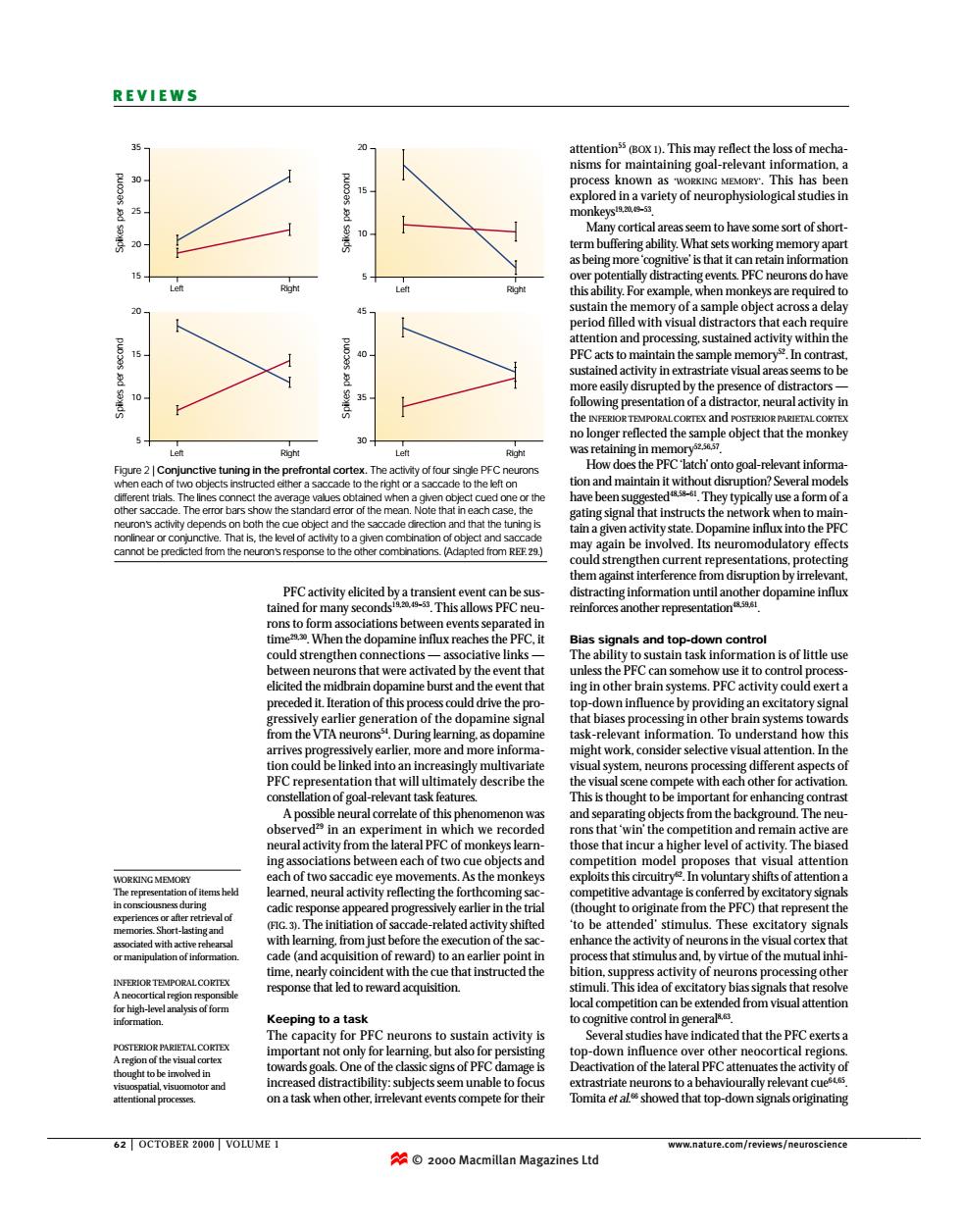
REVIEWS for ma ina variety of le ob ined activit bect tht the monk ont a-re ain it without disruption?Se ucts the en t olved.Its n uption by This allows PFC entation top-do control he eve of this pr ld dr ding an ons pr ing differer ought to be antorenherfot A po obs ed"in an in which we rec com e ar obiect KING MEMORY ooed )that represent th d)to spaergohe sual attention capacity for PFC neuro ns: the c signs of PF on of the teral PFCatte on a task when other ire events compete for the omita eal I that top-down signals originating 62 OCTOBER 2000 VOLUME 2000 Macmillan Magazines Lto 62 | OCTOBER 2000 | VOLUME 1 www.nature.com/reviews/neuroscience REVIEWS attention55 (BOX 1). This may reflect the loss of mechanisms for maintaining goal-relevant information, a process known as ‘WORKING MEMORY’. This has been explored in a variety of neurophysiological studies in monkeys19,20,49–53. Many cortical areas seem to have some sort of shortterm buffering ability. What sets working memory apart as being more ‘cognitive’ is that it can retain information over potentially distracting events. PFC neurons do have this ability. For example, when monkeys are required to sustain the memory of a sample object across a delay period filled with visual distractors that each require attention and processing, sustained activity within the PFC acts to maintain the sample memory52. In contrast, sustained activity in extrastriate visual areas seems to be more easily disrupted by the presence of distractors — following presentation of a distractor, neural activity in the INFERIOR TEMPORAL CORTEX and POSTERIOR PARIETAL CORTEX no longer reflected the sample object that the monkey was retaining in memory52,56,57. How does the PFC ‘latch’onto goal-relevant information and maintain it without disruption? Several models have been suggested48,58–61. They typically use a form of a gating signal that instructs the network when to maintain a given activity state. Dopamine influx into the PFC may again be involved. Its neuromodulatory effects could strengthen current representations, protecting them against interference from disruption by irrelevant, distracting information until another dopamine influx reinforces another representation48,59,61. Bias signals and top-down control The ability to sustain task information is of little use unless the PFC can somehow use it to control processing in other brain systems. PFC activity could exert a top-down influence by providing an excitatory signal that biases processing in other brain systems towards task-relevant information. To understand how this might work, consider selective visual attention. In the visual system, neurons processing different aspects of the visual scene compete with each other for activation. This is thought to be important for enhancing contrast and separating objects from the background. The neurons that ‘win’ the competition and remain active are those that incur a higher level of activity. The biased competition model proposes that visual attention exploits this circuitry62. In voluntary shifts of attention a competitive advantage is conferred by excitatory signals (thought to originate from the PFC) that represent the ‘to be attended’ stimulus. These excitatory signals enhance the activity of neurons in the visual cortex that process that stimulus and, by virtue of the mutual inhibition, suppress activity of neurons processing other stimuli. This idea of excitatory bias signals that resolve local competition can be extended from visual attention to cognitive control in general8,63. Several studies have indicated that the PFC exerts a top-down influence over other neocortical regions. Deactivation of the lateral PFC attenuates the activity of extrastriate neurons to a behaviourally relevant cue64,65. Tomita et al.66 showed that top-down signals originating PFC activity elicited by a transient event can be sustained for many seconds19,20,49–53. This allows PFC neurons to form associations between events separated in time29,30. When the dopamine influx reaches the PFC, it could strengthen connections — associative links — between neurons that were activated by the event that elicited the midbrain dopamine burst and the event that preceded it. Iteration of this process could drive the progressively earlier generation of the dopamine signal from the VTA neurons54. During learning, as dopamine arrives progressively earlier, more and more information could be linked into an increasingly multivariate PFC representation that will ultimately describe the constellation of goal-relevant task features. A possible neural correlate of this phenomenon was observed29 in an experiment in which we recorded neural activity from the lateral PFC of monkeys learning associations between each of two cue objects and each of two saccadic eye movements. As the monkeys learned, neural activity reflecting the forthcoming saccadic response appeared progressively earlier in the trial (FIG. 3). The initiation of saccade-related activity shifted with learning, from just before the execution of the saccade (and acquisition of reward) to an earlier point in time, nearly coincident with the cue that instructed the response that led to reward acquisition. Keeping to a task The capacity for PFC neurons to sustain activity is important not only for learning, but also for persisting towards goals. One of the classic signs of PFC damage is increased distractibility: subjects seem unable to focus on a task when other, irrelevant events compete for their WORKING MEMORY The representation of items held in consciousness during experiences or after retrieval of memories. Short-lasting and associated with active rehearsal or manipulation of information. INFERIOR TEMPORAL CORTEX A neocortical region responsible for high-level analysis of form information. POSTERIOR PARIETAL CORTEX A region of the visual cortex thought to be involved in visuospatial, visuomotor and attentional processes. 35 30 25 20 15 Left Spikes per second Right 20 15 10 5 Left Spikes per second Spikes per second Spikes per second Right 45 40 35 30 Left Right 20 15 10 5 Left Right Figure 2 | Conjunctive tuning in the prefrontal cortex. The activity of four single PFC neurons when each of two objects instructed either a saccade to the right or a saccade to the left on different trials. The lines connect the average values obtained when a given object cued one or the other saccade. The error bars show the standard error of the mean. Note that in each case, the neuron’s activity depends on both the cue object and the saccade direction and that the tuning is nonlinear or conjunctive. That is, the level of activity to a given combination of object and saccade cannot be predicted from the neuron’s response to the other combinations. (Adapted from REF. 29.) © 2000 Macmillan Magazines Ltd