正在加载图片...
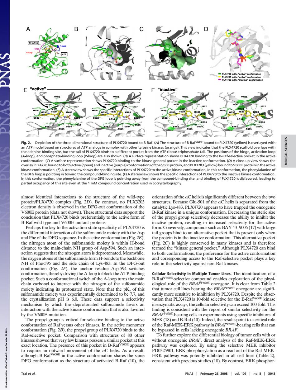
ctive (A)The stn n of this site even at the of the ntation of the oC heli rotein/P mplex(Fig ont no PL of the aC ted from th f ki a uniquc g the na protein,resulting inin the activ oups bind ket tha 20)is tive co in m al pocket. ough PLX4 can bine af-ective pocket playsa ky an r re Note that the of e are signifi mutation ding is con confor of Rafv hway in BRA aring cells that can 28).the Raf-selective cket.Co arison with structures of ot hou-Ra t n ERK path was DFG conformation as the structure of activated B-Raf(10),the consistent with previous studies (18).By contrast,ERK phosphor Tsai et al PNAS February 26.200 vol.105 no.04 almost identical interactions to the structure of the wild-type protein/PLX4720 complex (Fig. 2D). By contrast, no PLX3203 electron density is observed in the DFG-out conformation of the V600E protein (data not shown). These structural data support the conclusion that PLX4720 binds preferentially to the active form of B-Raf wild-type and V600E mutant proteins. Perhaps the key to the activation-state specificity of PLX4720 is the differential interaction of the sulfonamide moiety with the Asp and Phe of the DFG sequence. In the active conformation (Fig. 2E), the nitrogen atom of the sulfonamide moiety is within H-bond distance to the main-chain NH group of Asp-594. Such an interaction suggests that the nitrogen atom is deprotonated. Meanwhile, the oxygen atoms of the sulfonamide form H-bonds to the backbone NH of Phe-595 and the side chain of Lys-483. In the DFG-out conformation (Fig. 2F), the anchor residue Asp-594 switches conformation, thereby driving the A-loop to block the ATP-binding pocket. Such a conformational switch of the A-loop turns the main chain carbonyl to interact with the nitrogen of the sulfonamide moiety indicating its protonated state. Note that the pKa of this sulfonamide moiety was experimentally determined to be 7.7, and the crystallization pH is 6.0. These data support a selectivity mechanism by which the deprotonated sulfonamide favors an interaction with the active kinase conformation that is also favored by the V600E mutation. The propyl group is critical for selective binding to the active conformation of Raf versus other kinases. In the active monomer conformation (Fig. 2B), the propyl group of PLX4720 binds to the Raf-selective pocket. Comparison with structures of 80 other kinases showed that very few kinases possess a similar pocket at this exact location. The presence of this pocket in B-RafV600E appears to require an outward movement of the C helix. As a result, although B-RafV600E in the active conformation shares the same DFG conformation as the structure of activated B-Raf (10), the orientation of the C helix is significantly different between the two structures. Because Glu-501 of the C helix is separated from the catalytic Lys-483, PLX4720 appears to have trapped the oncogenic B-Raf kinase in a unique conformation. Decreasing the steric size of the propyl group selectively decreases the ability to inhibit the inactive protein, resulting in increased selectivity for the active form. Conversely, compounds such as BAY 43–9006 (17) with large tail groups bind to an alternative pocket that is present only when the protein is in the inactive conformation. This alternative pocket (Fig. 2C) is highly conserved in many kinases and is therefore termed the ‘‘kinase general pocket.’’ Although PLX4720 can bind to both conformations, the preference for the active conformation and corresponding access to the Raf-selective pocket plays a key role in the selectivity against non-Raf kinases. Cellular Selectivity in Multiple Tumor Lines. The identification of a B-RafV600E-selective compound enables exploration of the physiological role of the BRAFV600E oncogene. It is clear from Table 2 that tumor cell lines bearing the BRAFV600E oncogene are significantly more sensitive to inhibition by PLX4720. Despite the observation that PLX4720 is 10-fold selective for the B-RafV600E kinase in enzymatic assays, the cellular selectivity can exceed 100-fold. This finding is consistent with the report of similar selectivity for the BRAFV600E-bearing cells in experiments using specific inhibitors of MEK (18) and B-Raf (10). Indeed, the results point to a critical role of the Raf-MEK-ERK pathway in BRAFV600E-bearing cells that can be bypassed in cells lacking oncogenic BRAF. To further explore the differential biology of tumor cells with or without oncogenic BRAF, direct analysis of the Raf-MEK-ERK pathway was explored. By using the selective MEK inhibitor PD0325901, ERK phosphorylation as a read out of the Raf-MEKERK pathway was potently inhibited in all cell lines (Table 2), consistent with previous studies (18). By contrast, ERK phosphorPLX4720 ATP Hinge P-loop A-loop PLX4720 ATP Hinge P-loop A-loop “inactive” F583 F583 G593 G593 D594 D594 F595 F595 L514 L514 C532 C532 W531 W531 Q530 T529 Q530 T529 V471 V471 L505 L505 K483 K483 V482 V482 A481 A481 I527 I527 “inactive” F583 F583 G593 G593 D594 D594 F595 F595 L514 L514 C532 C532 W531 W531 Q530 T529 Q530 T529 V471 V471 L505 L505 K483 K483 V482 V482 A481 A481 I527 I527 F583 F583 G593 G593 D594 D594 F595 F595 L514 L514 C532 C532 W531 W531 Q530 T529 Q530 T529 V471 V471 L505 L505 K483 K483 V482 V482 A481 A481 I527 I527 “active” F583 F583 G593 G593 D594 D594 F595 F595 L514 L514 C532 C532 W531 W531 Q530 T529 Q530 T529 V471 V471 L505 L505 K483 K483 V482 V482 A481 A481 I527 I527 “active” C D E F PLX4720 in the “active” conformation PLX4720 in the “inactive” conformation PLX3203 in the “active” conformation PLX4720 in the “active” conformation PLX4720 in the “inactive” conformation PLX3203 in the “active” conformation A B Fig. 2. Depiction of the three-dimensional structure of PLX4720 bound to B-Raf. (A) The structure of B-RafV600E bound to PLX4720 (yellow) is overlayed with an ATP model based on structures of ATP analogs in complex with other tyrosine kinases (orange). This view indicates that the PLX4720 scaffold overlaps with the adenine-binding site, but the tail of PLX4720 binds to a different pocket from the ATP ribose-triphosphate tail. The positions of the hinge, activation loop (A-loop), and phosphate-binding loop (P-loop) are also shown. (B) A surface representation shows PLX4720 binding to the B-Raf-selective pocket in the active conformation. (C) A surface representation shows PLX4720 binding to the kinase general pocket in the inactive conformation. (D) A close-up view shows the overlay PLX4720 bound to both active (green) and inactive (purple) conformations of the V600 protein, and PLX3203 (yellow) bound to V600E protein in the active kinase conformation. (E) A stereoview shows the specific interactions of PLX4720 to the active kinase conformation. In this conformation, the phenylalanine of the DFG loop is pointing in toward the compound-binding site. (F) A stereoview shows the specific interactions of PLX4720 to the inactive kinase conformation. In this conformation, the phenylalanine of the DFG loop is pointing away from the compound-binding site, and binding of PLX4720 is disfavored, leading to partial occupancy of this site even at the 1 mM compound concentration used in cocrystallography. Tsai et al. PNAS February 26, 2008 vol. 105 no. 8 3043 MEDICAL SCIENCES Downloaded at Univ of Sci & Tech of China on December 23, 2019