正在加载图片...
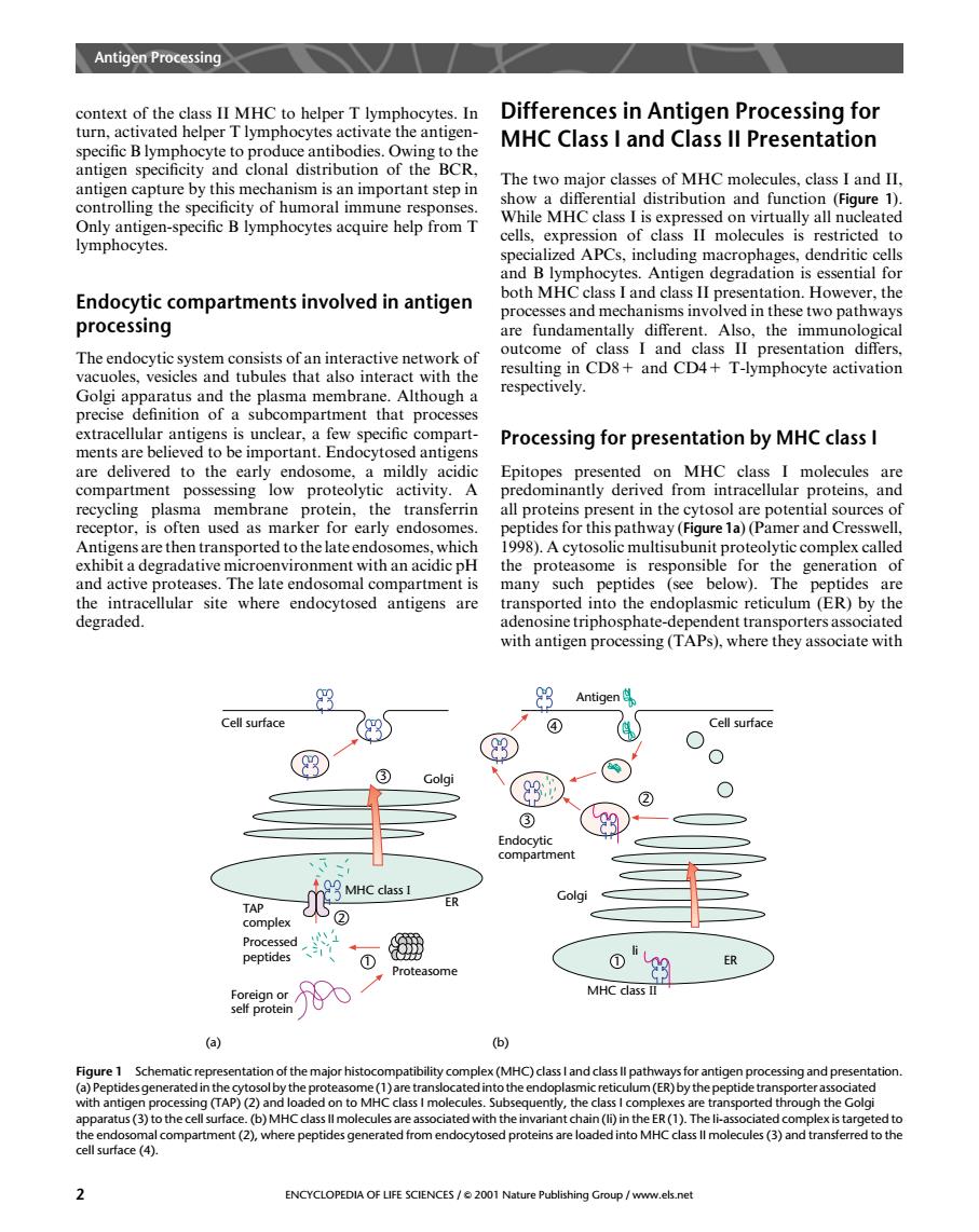
Antigen Proing context of the class II MHC to helper T lymphocytes.In Differences in Antigen Processing for turn,activated helper T lymphocytes activate the antigen- MHC Class I and Class ll Presentation gen spec The two maior classe s of MHC molecules,class I and II controlling the specificity of humoral immune resp 1 onses ntial distribut Only antigen-specific B lymphocytes acquire help from T Whil MHC casisprsdyceated lymphocytes. ng ma Endocytic compartments involved in antigen both MHC class I and class II pr entation.However.the processes and mechanisms involved in these two pathways processing are fundamentally different presenta ctiv precise Processing for presentation by MHC class I ed to the mildly A FpicinPiyaedeamMHSdilhrpoas MHC clas molecules recycling plasma membrane all proteins present in the cytosol are potential sources receptor. ten used as n early endos s for thi (Figure a)(P and C ansported t tan acidic pH the .A cyto: me and active proteases.The late endosomal compartment is transported into the endoplasmic reticulum(ER)by the with antigen processing(T they associate with Antigen Cell surface Cell surface Golgi TAP 2 peptide Proteasome 0"9 MHC dass II histo (MHC) us(3)to the cell su (b)MHC cass ll mol ted with thei n(li)in the ER(1).T t(2).where pep s genera s (3)and tran ENCYCLOPEDIA OF LIFE SCIENCES/e 2001 Nature Publishing Group /www.els.netcontext of the class II MHC to helper T lymphocytes. In turn, activated helper T lymphocytes activate the antigenspecific B lymphocyte to produce antibodies. Owing to the antigen specificity and clonal distribution of the BCR, antigen capture by this mechanism is an important step in controlling the specificity of humoral immune responses. Only antigen-specific B lymphocytes acquire help from T lymphocytes. Endocytic compartments involved in antigen processing The endocytic system consists of an interactive network of vacuoles, vesicles and tubules that also interact with the Golgi apparatus and the plasma membrane. Although a precise definition of a subcompartment that processes extracellular antigens is unclear, a few specific compartments are believed to be important. Endocytosed antigens are delivered to the early endosome, a mildly acidic compartment possessing low proteolytic activity. A recycling plasma membrane protein, the transferrin receptor, is often used as marker for early endosomes. Antigens are then transported to the late endosomes, which exhibit a degradative microenvironment with an acidic pH and active proteases. The late endosomal compartment is the intracellular site where endocytosed antigens are degraded. Differences in Antigen Processing for MHC Class I and Class II Presentation The two major classes of MHC molecules, class I and II, show a differential distribution and function (Figure 1). While MHC class I is expressed on virtually all nucleated cells, expression of class II molecules is restricted to specialized APCs, including macrophages, dendritic cells and B lymphocytes. Antigen degradation is essential for both MHC class I and class II presentation. However, the processes and mechanisms involved in these two pathways are fundamentally different. Also, the immunological outcome of class I and class II presentation differs, resulting in CD8+ and CD4+ T-lymphocyte activation respectively. Processing for presentation by MHC class I Epitopes presented on MHC class I molecules are predominantly derived from intracellular proteins, and all proteins present in the cytosol are potential sources of peptides for this pathway (Figure 1a) (Pamer and Cresswell, 1998). A cytosolic multisubunit proteolytic complex called the proteasome is responsible for the generation of many such peptides (see below). The peptides are transported into the endoplasmic reticulum (ER) by the adenosine triphosphate-dependent transporters associated with antigen processing (TAPs), where they associate with (a) (b) Cell surface 3 Golgi MHC class I 2 TAP complex Processed peptides ER Proteasome 1 Foreign or self protein Antigen 4 MHC class II 2 Endocytic compartment 1 ER li 3 Cell surface Golgi Figure 1 Schematic representation of the major histocompatibility complex (MHC) class I and class II pathways for antigen processing and presentation. (a) Peptides generated in the cytosolby the proteasome (1)are translocated into the endoplasmicreticulum (ER) by the peptide transporter associated with antigen processing (TAP) (2) and loaded on to MHC class I molecules. Subsequently, the class I complexes are transported through the Golgi apparatus (3) to the cell surface. (b) MHC class II molecules are associated with the invariant chain (Ii) in the ER (1). The Ii-associated complex is targeted to the endosomal compartment (2), where peptides generated from endocytosed proteins are loaded into MHC class II molecules (3) and transferred to the cell surface (4). Antigen Processing 2 ENCYCLOPEDIA OF LIFE SCIENCES / & 2001 Nature Publishing Group / www.els.net