正在加载图片...
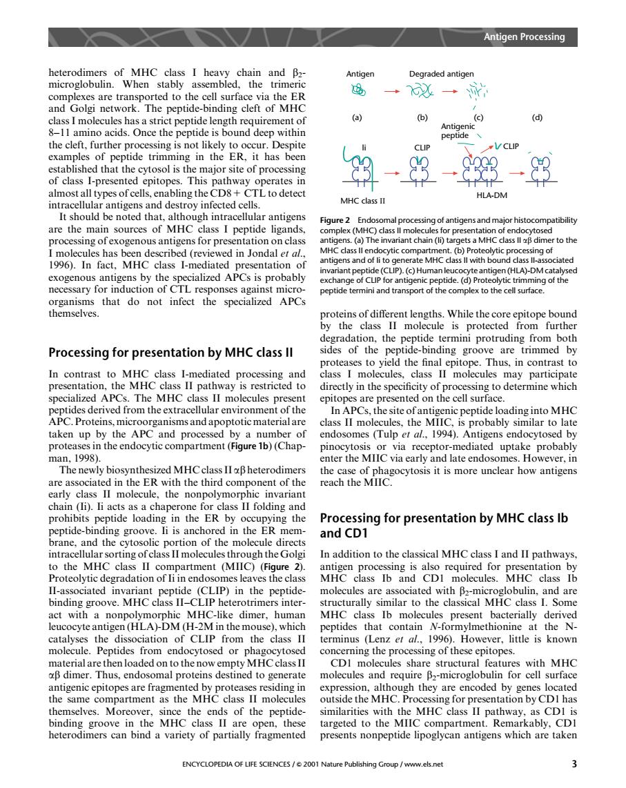
Antigen Processing Antigen Degraded antigen rted to th surf a the ER and Golgi network.The peptide-binding cleft of MHC b) (c) the clen. Onc the peptide dcep with nthe ER.ith CLIP of class I-presented epitope This pathway operates I almost lI types of cel ling the CD8- HLA-DM MHC class II antigens are the main sources of MHC class I peptide ligands, Figure2 MHC ant chai o the 1996.1n ate cialized APCs is probably UP) eptide terminiand tran ves e epitope bound the Processing for presentation by MHC class ll sides of the peptide-binding groove are trimmed by proteases to yield the final epitope.Thus,in contrast to cule ass II molecules ialized APCs The MHC class I r peptides derived from the extracellular envromenofthe In aPCs the siteofant peptide loading into MHC s II mole by the es,the MI bably similar to late and pro s(Tulp et al. 1994 Ant r in The newly biosynthesized MHCclassIIBheterodimers the case of phagocytosis it is more unclear how antigens reach the MIIC n (i). the nonpo prohibits。 eptide loading in the ER by o ing the Processing for presentation by MHC class Ib clnd the and CD1 portion of the mo In addition to the elassical MHCclass I and II path vays MHC class Ib and CDI molecules.MHC class Ib Hl-associated invariant peptide (CLIP)in the peptide- molecules are associated with B2-microglobulin,and are binding groove.MHC trimers inter milar to th classical M 1.S0m act witl dimer id cla catalyses the dissociation of CLIP from the class II terminus (Lenz).However.lite is known molecule.Peptides from endocytosed or pha concerning the processing of these epitopes. hen lo aded on to the now sdiengMhiCchasi CDI mole ecules share structura en nal pro p2-micro h th the same compartment as the MHc class molecules outside the MHC Processine for presentation by CDIhas similarities with the MHC class II pathway,as CDI is ners can t a variety class n presents nonpeptide lipoglycan antigens w are taken ENCYCLOPEDIA OF LIFE SCIENCES/2001 Nature Publishing Group/www.els.net heterodimers of MHC class I heavy chain and b2- microglobulin. When stably assembled, the trimeric complexes are transported to the cell surface via the ER and Golgi network. The peptide-binding cleft of MHC class I molecules has a strict peptide length requirement of 8–11 amino acids. Once the peptide is bound deep within the cleft, further processing is not likely to occur. Despite examples of peptide trimming in the ER, it has been established that the cytosol is the major site of processing of class I-presented epitopes. This pathway operates in almost all types of cells, enabling the CD8+ CTL to detect intracellular antigens and destroy infected cells. It should be noted that, although intracellular antigens are the main sources of MHC class I peptide ligands, processing of exogenous antigens for presentation on class I molecules has been described (reviewed in Jondal et al., 1996). In fact, MHC class I-mediated presentation of exogenous antigens by the specialized APCs is probably necessary for induction of CTL responses against microorganisms that do not infect the specialized APCs themselves. Processing for presentation by MHC class II In contrast to MHC class I-mediated processing and presentation, the MHC class II pathway is restricted to specialized APCs. The MHC class II molecules present peptides derived from the extracellular environment of the APC. Proteins, microorganisms and apoptotic material are taken up by the APC and processed by a number of proteases in the endocytic compartment (Figure 1b) (Chapman, 1998). The newly biosynthesizedMHC class II ab heterodimers are associated in the ER with the third component of the early class II molecule, the nonpolymorphic invariant chain (Ii). Ii acts as a chaperone for class II folding and prohibits peptide loading in the ER by occupying the peptide-binding groove. Ii is anchored in the ER membrane, and the cytosolic portion of the molecule directs intracellular sorting of class II molecules through the Golgi to the MHC class II compartment (MIIC) (Figure 2). Proteolytic degradation of Ii in endosomes leaves the class II-associated invariant peptide (CLIP) in the peptidebinding groove. MHC class II–CLIP heterotrimers interact with a nonpolymorphic MHC-like dimer, human leucocyte antigen (HLA)-DM (H-2M in the mouse), which catalyses the dissociation of CLIP from the class II molecule. Peptides from endocytosed or phagocytosed material are then loaded on to the now emptyMHC class II ab dimer. Thus, endosomal proteins destined to generate antigenic epitopes are fragmented by proteases residing in the same compartment as the MHC class II molecules themselves. Moreover, since the ends of the peptidebinding groove in the MHC class II are open, these heterodimers can bind a variety of partially fragmented proteins of different lengths. While the core epitope bound by the class II molecule is protected from further degradation, the peptide termini protruding from both sides of the peptide-binding groove are trimmed by proteases to yield the final epitope. Thus, in contrast to class I molecules, class II molecules may participate directly in the specificity of processing to determine which epitopes are presented on the cell surface. In APCs, the site of antigenic peptide loading into MHC class II molecules, the MIIC, is probably similar to late endosomes (Tulp et al., 1994). Antigens endocytosed by pinocytosis or via receptor-mediated uptake probably enter the MIIC via early and late endosomes. However, in the case of phagocytosis it is more unclear how antigens reach the MIIC. Processing for presentation by MHC class Ib and CD1 In addition to the classical MHC class I and II pathways, antigen processing is also required for presentation by MHC class Ib and CD1 molecules. MHC class Ib molecules are associated with b2-microglobulin, and are structurally similar to the classical MHC class I. Some MHC class Ib molecules present bacterially derived peptides that contain N-formylmethionine at the Nterminus (Lenz et al., 1996). However, little is known concerning the processing of these epitopes. CD1 molecules share structural features with MHC molecules and require b2-microglobulin for cell surface expression, although they are encoded by genes located outside the MHC. Processing for presentation by CD1 has similarities with the MHC class II pathway, as CD1 is targeted to the MIIC compartment. Remarkably, CD1 presents nonpeptide lipoglycan antigens which are taken MHC class II li (a) CLIP (b) Antigenic peptide CLIP HLA-DM (c) (d) Antigen Degraded antigen Figure 2 Endosomal processing of antigens and major histocompatibility complex (MHC) class II molecules for presentation of endocytosed antigens. (a) The invariant chain (Ii) targets a MHC class II ab dimer to the MHC class II endocytic compartment. (b) Proteolytic processing of antigens and of Ii to generate MHC class II with bound class II-associated invariant peptide (CLIP). (c) Human leucocyte antigen (HLA)-DM catalysed exchange of CLIP for antigenic peptide. (d) Proteolytic trimming of the peptide termini and transport of the complex to the cell surface. Antigen Processing ENCYCLOPEDIA OF LIFE SCIENCES / & 2001 Nature Publishing Group / www.els.net 3