正在加载图片...
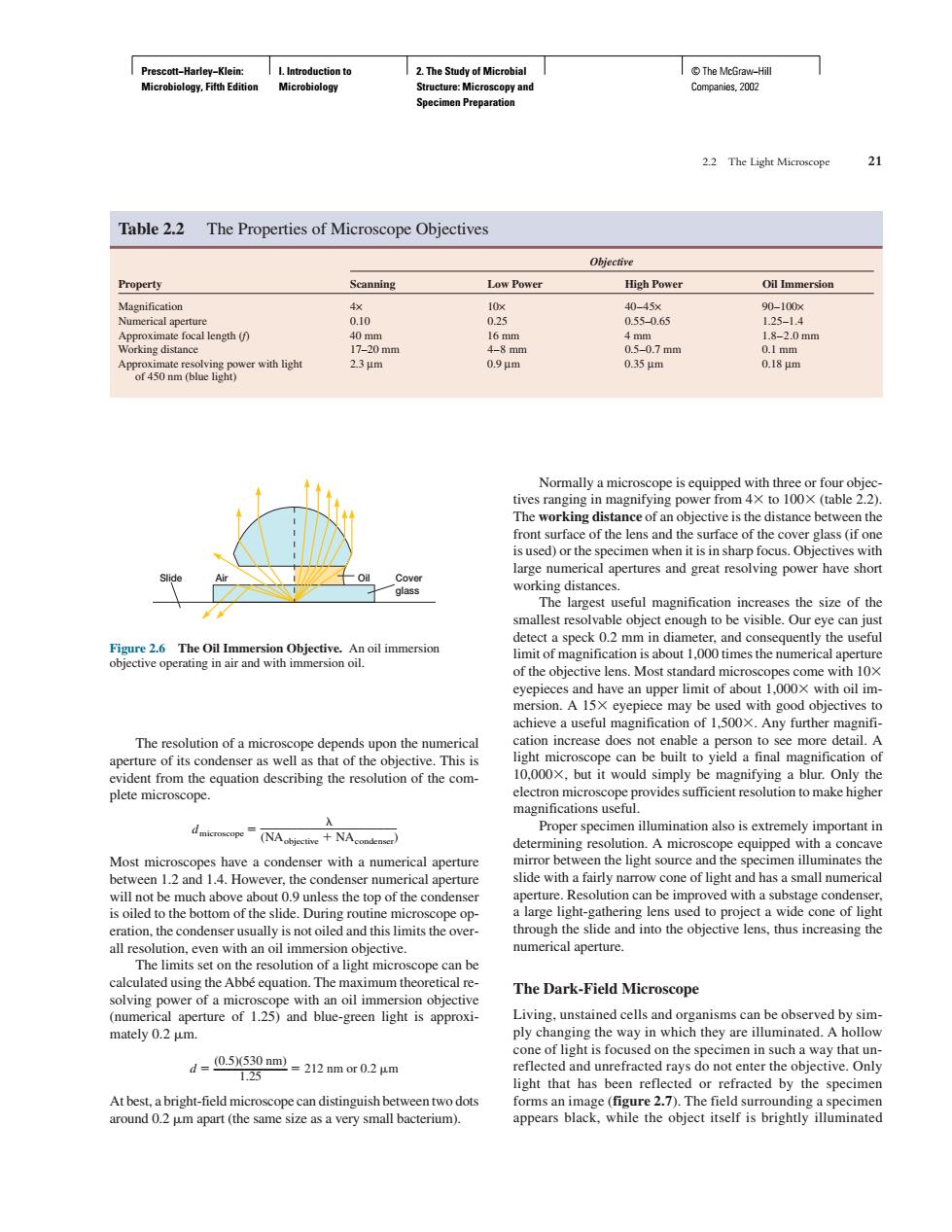
2.The Study Microbial Table 2.2 The Properties of Microscope Objectives Low Powt High Powe 00 035 Normally a micre cope is equipped with three or four front surface of the lens and the surface of the cover glass (if on The and cor mes the n achieve a useful magnification of 1500.Any further ma The resolution of a microscope depends upon the numerica 10.000x,but it would simply be magnifying a blur.Only the Proper specimen illumination also is extremely imp tant in th a slide with a fairly narow con of lighta has a small nur ncrica numerical aperture The Dark-Field Microscope Living.unstained cells and organis sms can be observed by sim mately 0.2 um. ply cha 4=0550里-2m02 reflected and unrefracte d rays do not enter the ob ight that has be en retl the specimePrescott−Harley−Klein: Microbiology, Fifth Edition I. Introduction to Microbiology 2. The Study of Microbial Structure: Microscopy and Specimen Preparation © The McGraw−Hill Companies, 2002 The resolution of a microscope depends upon the numerical aperture of its condenser as well as that of the objective. This is evident from the equation describing the resolution of the complete microscope. dmicroscope ______________________ (NAobjective NAcondenser) Most microscopes have a condenser with a numerical aperture between 1.2 and 1.4. However, the condenser numerical aperture will not be much above about 0.9 unless the top of the condenser is oiled to the bottom of the slide. During routine microscope operation, the condenser usually is not oiled and this limits the overall resolution, even with an oil immersion objective. The limits set on the resolution of a light microscope can be calculated using the Abbé equation. The maximum theoretical resolving power of a microscope with an oil immersion objective (numerical aperture of 1.25) and blue-green light is approximately 0.2 m. (0.5)(530 nm) d –––––––––––– 212 nm or 0.2 m 1.25 At best, a bright-field microscope can distinguish between two dots around 0.2 m apart (the same size as a very small bacterium). Normally a microscope is equipped with three or four objectives ranging in magnifying power from 4 to 100 (table 2.2). The working distance of an objective is the distance between the front surface of the lens and the surface of the cover glass (if one is used) or the specimen when it is in sharp focus. Objectives with large numerical apertures and great resolving power have short working distances. The largest useful magnification increases the size of the smallest resolvable object enough to be visible. Our eye can just detect a speck 0.2 mm in diameter, and consequently the useful limit of magnification is about 1,000 times the numerical aperture of the objective lens. Most standard microscopes come with 10 eyepieces and have an upper limit of about 1,000 with oil immersion. A 15 eyepiece may be used with good objectives to achieve a useful magnification of 1,500. Any further magnification increase does not enable a person to see more detail. A light microscope can be built to yield a final magnification of 10,000, but it would simply be magnifying a blur. Only the electron microscope provides sufficient resolution to make higher magnifications useful. Proper specimen illumination also is extremely important in determining resolution. A microscope equipped with a concave mirror between the light source and the specimen illuminates the slide with a fairly narrow cone of light and has a small numerical aperture. Resolution can be improved with a substage condenser, a large light-gathering lens used to project a wide cone of light through the slide and into the objective lens, thus increasing the numerical aperture. The Dark-Field Microscope Living, unstained cells and organisms can be observed by simply changing the way in which they are illuminated. A hollow cone of light is focused on the specimen in such a way that unreflected and unrefracted rays do not enter the objective. Only light that has been reflected or refracted by the specimen forms an image (figure 2.7). The field surrounding a specimen appears black, while the object itself is brightly illuminated 2.2 The Light Microscope 21 Table 2.2 The Properties of Microscope Objectives Objective Property Scanning Low Power High Power Oil Immersion Magnification 4× 10× 40–45× 90–100× Numerical aperture 0.10 0.25 0.55–0.65 1.25–1.4 Approximate focal length (f) 40 mm 16 mm 4 mm 1.8–2.0 mm Working distance 17–20 mm 4–8mm 0.5–0.7 mm 0.1 mm Approximate resolving power with light 2.3 µm 0.9 µm 0.35 µm 0.18 µm of 450 nm (blue light) Air Oil Cover glass Slide Figure 2.6 The Oil Immersion Objective. An oil immersion objective operating in air and with immersion oil