正在加载图片...
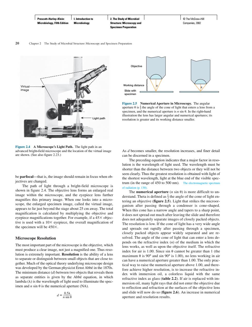
。 pter 2 Figure 25 Numerical Aperture in Micr nd t 6.In ti resolution is greater and its working distance smaller Figure 24 A Micn As d becomes smaller.the resolution increases.and finer detail re shown (See also figure 2.23.) can be disceme ed in a specimen the shortest waveler light at the blueend of the visible spec tum (in the range of 0 t0 500 m). image within the microsc ope.and the eyepiece lens further this primary imge When one out 25 cm aw y.The to spreads out sin througha specimen clos Microscope Resolution ed packed ds on the refractive index (n)of the mediun in which the The most important part of the microscope is the objective.which .Thus reso upon the objective itself. Th musprotuceaClerimage,notiLa small objects that are imum0is90°and sin90°isL.00). ng in air to s to rais fore achieve higher res tion.i toincrease the refractive in imen andnsin is the numerical aperture (NA). d-品 and slide will now do so(figure 2.6).An increase in numerical aperture and resolution results. Prescott−Harley−Klein: Microbiology, Fifth Edition I. Introduction to Microbiology 2. The Study of Microbial Structure: Microscopy and Specimen Preparation © The McGraw−Hill Companies, 2002 be parfocal—that is, the image should remain in focus when objectives are changed. The path of light through a bright-field microscope is shown in figure 2.4. The objective lens forms an enlarged real image within the microscope, and the eyepiece lens further magnifies this primary image. When one looks into a microscope, the enlarged specimen image, called the virtual image, appears to lie just beyond the stage about 25 cm away. The total magnification is calculated by multiplying the objective and eyepiece magnifications together. For example, if a 45 objective is used with a 10 eyepiece, the overall magnification of the specimen will be 450. Microscope Resolution The most important part of the microscope is the objective, which must produce a clear image, not just a magnified one. Thus resolution is extremely important. Resolution is the ability of a lens to separate or distinguish between small objects that are close together. Much of the optical theory underlying microscope design was developed by the German physicist Ernst Abbé in the 1870s. The minimum distance (d) between two objects that reveals them as separate entities is given by the Abbé equation, in which lambda () is the wavelength of light used to illuminate the specimen and n sin is the numerical aperture (NA). 0.5 d _____ n sin As d becomes smaller, the resolution increases, and finer detail can be discerned in a specimen. The preceding equation indicates that a major factor in resolution is the wavelength of light used. The wavelength must be shorter than the distance between two objects or they will not be seen clearly. Thus the greatest resolution is obtained with light of the shortest wavelength, light at the blue end of the visible spectrum (in the range of 450 to 500 nm). The electromagnetic spectrum of radiation (p. 130). The numerical aperture (n sin ) is more difficult to understand. Theta is defined as 1 2 the angle of the cone of light entering an objective (figure 2.5). Light that strikes the microorganism after passing through a condenser is cone-shaped. When this cone has a narrow angle and tapers to a sharp point, it does not spread out much after leaving the slide and therefore does not adequately separate images of closely packed objects. The resolution is low. If the cone of light has a very wide angle and spreads out rapidly after passing through a specimen, closely packed objects appear widely separated and are resolved. The angle of the cone of light that can enter a lens depends on the refractive index (n) of the medium in which the lens works, as well as upon the objective itself. The refractive index for air is 1.00. Since sin cannot be greater than 1 (the maximum is 90° and sin 90° is 1.00), no lens working in air can have a numerical aperture greater than 1.00. The only practical way to raise the numerical aperture above 1.00, and therefore achieve higher resolution, is to increase the refractive index with immersion oil, a colorless liquid with the same refractive index as glass (table 2.2). If air is replaced with immersion oil, many light rays that did not enter the objective due to reflection and refraction at the surfaces of the objective lens and slide will now do so (figure 2.6). An increase in numerical aperture and resolution results. 20 Chapter 2 The Study of Microbial Structure: Microscopy and Specimen Preparation Light path Figure 2.4 A Microscope’s Light Path. The light path in an advanced bright-field microscope and the location of the virtual image are shown. (See also figure 2.23.) Objective Working distance Slide with specimen θ θ Figure 2.5 Numerical Aperture in Microscopy. The angular aperture is 1 2 the angle of the cone of light that enters a lens from a specimen, and the numerical aperture is n sin . In the right-hand illustration the lens has larger angular and numerical apertures; its resolution is greater and its working distance smaller.�������