正在加载图片...
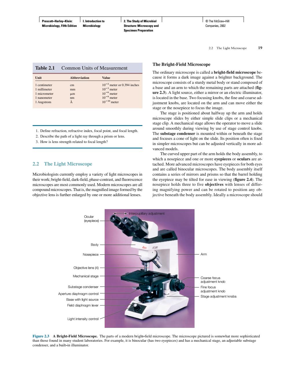
19 Table 2.1 Common Units of Measurement The Bright-Field Microscope -field mic Unit Abbreviation ure2.3).A light source.either amirororanelectric illuminat stage clip.A chanical stage a s the operator to mo 1.Define refraction,refractive index.focal point,and focal length substag conde r is mo unted withinor beneath the a cone of lightont e sh which 2.2 The Light Microscope tached.More contains a series of mirrors and prisms so that the barrel holding ing magnifying power and can be rotated to position any ob- objective lens is further enlaged by oneor more additional lenses. jective beneath the body assembly.Ideally a microscope should Body Nosepiece Obiective lens (4) Mechanical sge Apertu r diaphragm control nt knob Bright-Field Mie scope.Prescott−Harley−Klein: Microbiology, Fifth Edition I. Introduction to Microbiology 2. The Study of Microbial Structure: Microscopy and Specimen Preparation © The McGraw−Hill Companies, 2002 1. Define refraction, refractive index, focal point, and focal length. 2. Describe the path of a light ray through a prism or lens. 3. How is lens strength related to focal length? 2.2 The Light Microscope Microbiologists currently employ a variety of light microscopes in their work; bright-field, dark-field, phase-contrast, and fluorescence microscopes are most commonly used. Modern microscopes are all compound microscopes. That is, the magnified image formed by the objective lens is further enlarged by one or more additional lenses. The Bright-Field Microscope The ordinary microscope is called a bright-field microscope because it forms a dark image against a brighter background. The microscope consists of a sturdy metal body or stand composed of a base and an arm to which the remaining parts are attached (figure 2.3). A light source, either a mirror or an electric illuminator, is located in the base. Two focusing knobs, the fine and coarse adjustment knobs, are located on the arm and can move either the stage or the nosepiece to focus the image. The stage is positioned about halfway up the arm and holds microscope slides by either simple slide clips or a mechanical stage clip. A mechanical stage allows the operator to move a slide around smoothly during viewing by use of stage control knobs. The substage condenser is mounted within or beneath the stage and focuses a cone of light on the slide. Its position often is fixed in simpler microscopes but can be adjusted vertically in more advanced models. The curved upper part of the arm holds the body assembly, to which a nosepiece and one or more eyepieces or oculars are attached. More advanced microscopes have eyepieces for both eyes and are called binocular microscopes. The body assembly itself contains a series of mirrors and prisms so that the barrel holding the eyepiece may be tilted for ease in viewing (figure 2.4). The nosepiece holds three to five objectives with lenses of differing magnifying power and can be rotated to position any objective beneath the body assembly. Ideally a microscope should 2.2 The Light Microscope 19 Table 2.1 Common Units of Measurement Unit Abbreviation Value 1 centimeter cm 102 meter or 0.394 inches 1 millimeter mm 103 meter 1 micrometer m 106 meter 1 nanometer nm 109 meter 1 Angstrom Å 1010 meter Ocular (eyepiece) Body Arm Coarse focus adjustment knob Fine focus adjustment knob Stage adjustment knobs Interpupillary adjustment Nosepiece Objective lens (4) Mechanical stage Substage condenser Aperture diaphragm control Base with light source Field diaphragm lever Light intensity control Figure 2.3 A Bright-Field Microscope. The parts of a modern bright-field microscope. The microscope pictured is somewhat more sophisticated than those found in many student laboratories. For example, it is binocular (has two eyepieces) and has a mechanical stage, an adjustable substage condenser, and a built-in illuminator.�����