正在加载图片...
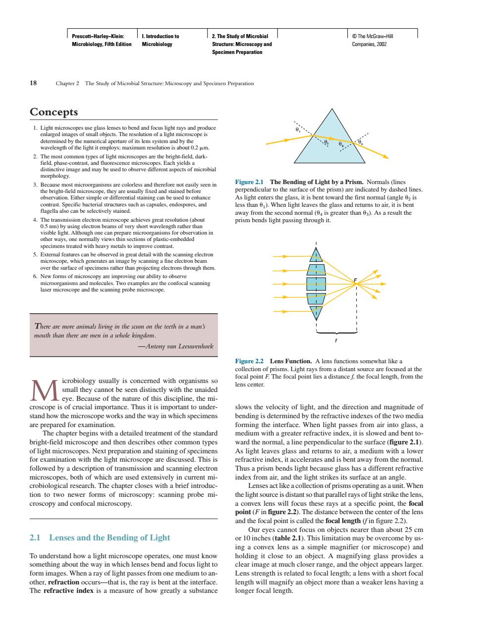
8 Concepts of its len s system a nd by the 2 opboloey Figure2 The Bending of Light bya Prism.Nomals (ines (o are more animals living in the scum on the tocth in a man's nouth than there are men in a whole kingdom. /Antony yn Leeuwenhock d at the M Be ial in s the micros nitude o works and the way in which specimens the refractive indexes of the two media bright-field microscope and then describes other common types ward the normal,a line perpendicular to the surface(figure 2.1) Thus a prism bends light e glass has a different refractive croscopy:scanning probe mi istant so that parallel rays of light strike the e focal point is called the fo length (in figure 2.1 Lenses and the Bending of Light something ab clear image at much close c,and the appears large othemere of ow longer focal length Prescott−Harley−Klein: Microbiology, Fifth Edition I. Introduction to Microbiology 2. The Study of Microbial Structure: Microscopy and Specimen Preparation © The McGraw−Hill Companies, 2002 Concepts 1. Light microscopes use glass lenses to bend and focus light rays and produce enlarged images of small objects. The resolution of a light microscope is determined by the numerical aperture of its lens system and by the wavelength of the light it employs; maximum resolution is about 0.2 m. 2. The most common types of light microscopes are the bright-field, darkfield, phase-contrast, and fluorescence microscopes. Each yields a distinctive image and may be used to observe different aspects of microbial morphology. 3. Because most microorganisms are colorless and therefore not easily seen in the bright-field microscope, they are usually fixed and stained before observation. Either simple or differential staining can be used to enhance contrast. Specific bacterial structures such as capsules, endospores, and flagella also can be selectively stained. 4. The transmission electron microscope achieves great resolution (about 0.5 nm) by using electron beams of very short wavelength rather than visible light. Although one can prepare microorganisms for observation in other ways, one normally views thin sections of plastic-embedded specimens treated with heavy metals to improve contrast. 5. External features can be observed in great detail with the scanning electron microscope, which generates an image by scanning a fine electron beam over the surface of specimens rather than projecting electrons through them. 6. New forms of microscopy are improving our ability to observe microorganisms and molecules. Two examples are the confocal scanning laser microscope and the scanning probe microscope. There are more animals living in the scum on the teeth in a man’s mouth than there are men in a whole kingdom. —Antony van Leeuwenhoek Microbiology usually is concerned with organisms so small they cannot be seen distinctly with the unaided eye. Because of the nature of this discipline, the microscope is of crucial importance. Thus it is important to understand how the microscope works and the way in which specimens are prepared for examination. The chapter begins with a detailed treatment of the standard bright-field microscope and then describes other common types of light microscopes. Next preparation and staining of specimens for examination with the light microscope are discussed. This is followed by a description of transmission and scanning electron microscopes, both of which are used extensively in current microbiological research. The chapter closes with a brief introduction to two newer forms of microscopy: scanning probe microscopy and confocal microscopy. 2.1 Lenses and the Bending of Light To understand how a light microscope operates, one must know something about the way in which lenses bend and focus light to form images. When a ray of light passes from one medium to another, refraction occurs—that is, the ray is bent at the interface. The refractive index is a measure of how greatly a substance 18 Chapter 2 The Study of Microbial Structure: Microscopy and Specimen Preparation slows the velocity of light, and the direction and magnitude of bending is determined by the refractive indexes of the two media forming the interface. When light passes from air into glass, a medium with a greater refractive index, it is slowed and bent toward the normal, a line perpendicular to the surface (figure 2.1). As light leaves glass and returns to air, a medium with a lower refractive index, it accelerates and is bent away from the normal. Thus a prism bends light because glass has a different refractive index from air, and the light strikes its surface at an angle. Lenses act like a collection of prisms operating as a unit. When the light source is distant so that parallel rays of light strike the lens, a convex lens will focus these rays at a specific point, the focal point (F in figure 2.2). The distance between the center of the lens and the focal point is called the focal length (f in figure 2.2). Our eyes cannot focus on objects nearer than about 25 cm or 10 inches (table 2.1). This limitation may be overcome by using a convex lens as a simple magnifier (or microscope) and holding it close to an object. A magnifying glass provides a clear image at much closer range, and the object appears larger. Lens strength is related to focal length; a lens with a short focal length will magnify an object more than a weaker lens having a longer focal length. 4 3 2 1 θ θ θ θ Figure 2.1 The Bending of Light by a Prism. Normals (lines perpendicular to the surface of the prism) are indicated by dashed lines. As light enters the glass, it is bent toward the first normal (angle 2 is less than 1). When light leaves the glass and returns to air, it is bent away from the second normal (4 is greater than 3). As a result the prism bends light passing through it. f F Figure 2.2 Lens Function. A lens functions somewhat like a collection of prisms. Light rays from a distant source are focused at the focal point F. The focal point lies a distance f, the focal length, from the lens center.����