正在加载图片...
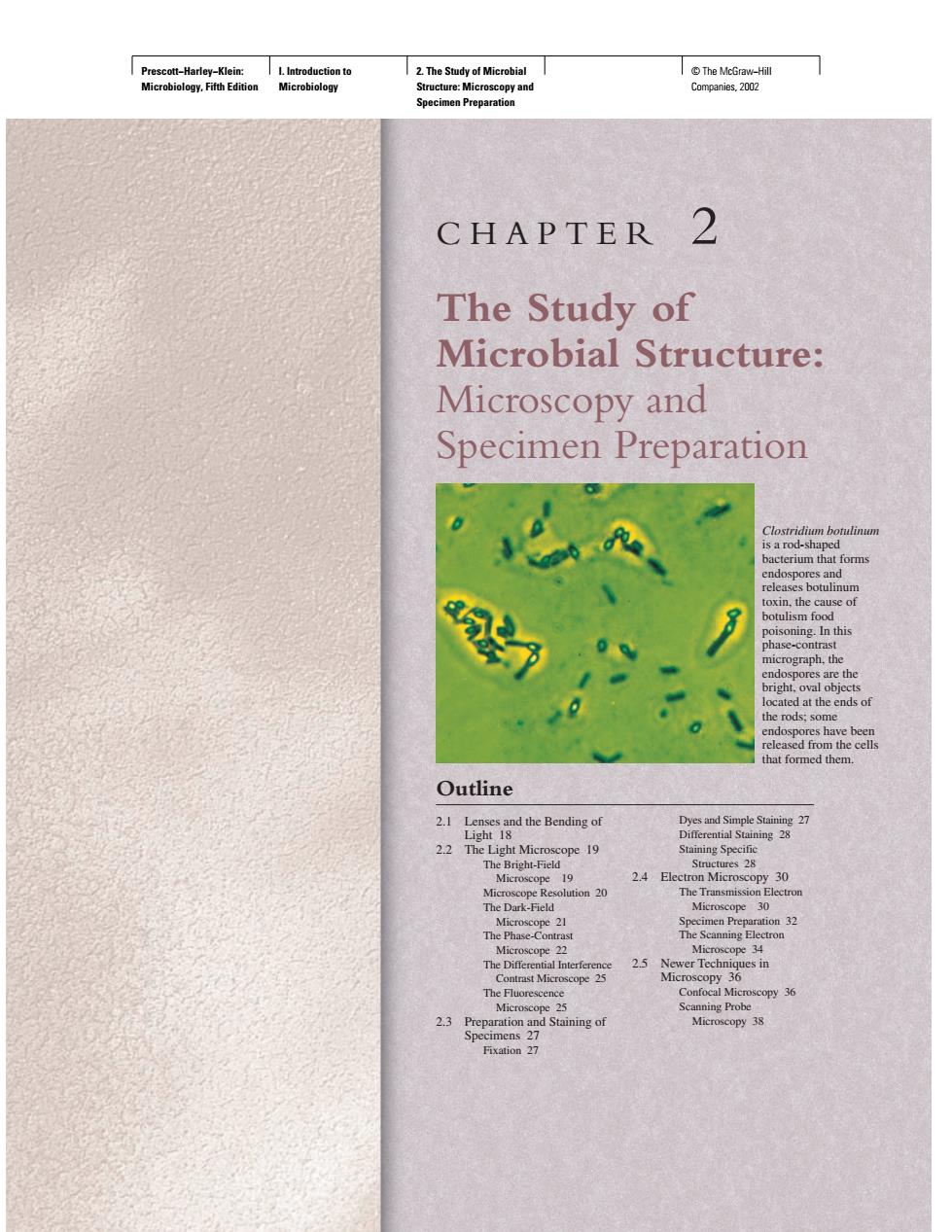
CHAPTER 2 The Study of Microbial Structure: Microscopy and Specimen Preparation at the rods,so ed from the cell at formed ther Outline 21e the Bending of 22 Th cope 19 Brigh-F 19 24日 30 25 Newer Techni 3 reparation Microscopy 38Prescott−Harley−Klein: Microbiology, Fifth Edition I. Introduction to Microbiology 2. The Study of Microbial Structure: Microscopy and Specimen Preparation © The McGraw−Hill Companies, 2002 CHAPTER 2 The Study of Microbial Structure: Microscopy and Specimen Preparation Clostridium botulinum is a rod-shaped bacterium that forms endospores and releases botulinum toxin, the cause of botulism food poisoning. In this phase-contrast micrograph, the endospores are the bright, oval objects located at the ends of the rods; some endospores have been released from the cells that formed them. 2.1 Lenses and the Bending of Light 18 2.2 The Light Microscope 19 The Bright-Field Microscope 19 Microscope Resolution 20 The Dark-Field Microscope 21 The Phase-Contrast Microscope 22 The Differential Interference Contrast Microscope 25 The Fluorescence Microscope 25 2.3 Preparation and Staining of Specimens 27 Fixation 27 Dyes and Simple Staining 27 Differential Staining 28 Staining Specific Structures 28 2.4 Electron Microscopy 30 The Transmission Electron Microscope 30 Specimen Preparation 32 The Scanning Electron Microscope 34 2.5 Newer Techniques in Microscopy 36 Confocal Microscopy 36 Scanning Probe Microscopy 38 Outline