正在加载图片...
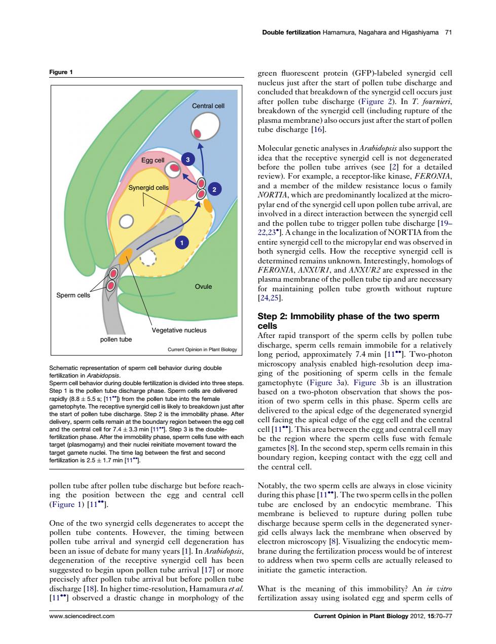
Double fertilization Hamamura.Nagahara and Higashivama 71 Figure 1 e and concluded that breakdown of the synergid cell occurs just afterolenowfth dischar In T. fe theopn Molecular genetic analyses in Arabidopsis also support the erated r-like 2l for mber of the m二com nich are pre at the micro charge [19- zation or plasma membrane of the pollen tube tip and are nccessary Sperm cells ning pllen tubethupre Step 2:Immobility phase of the two sperm t of the oollen tube long period,approximately 7.4 min [11".Two-photon cell be havior during double in ana alysis enab high-re ametophyte (Figure 3a).Figure 3b is an illustration ase d on a two-photor nthat shows the pos dclivered to the apical cdge of the svnerid tep 2 is th ng the apical edge of the egg cel and the central This area pthe I ma th fe he cent pollen tube a rm c d b membrane is believed to rupture during pollen tube ccept the pollen tube arrival and synergid cell de neration has clectron microscop Visu the ate for many years [ idopst recep the a s are actually re preciscly after am a drasti www.sciencedirect.com Current Opinion in Plant Biology 0.1:70-77pollen tube after pollen tube discharge but before reaching the position between the egg and central cell (Figure 1) [11]. One of the two synergid cells degenerates to accept the pollen tube contents. However, the timing between pollen tube arrival and synergid cell degeneration has been an issue of debate for many years [1]. In Arabidopsis, degeneration of the receptive synergid cell has been suggested to begin upon pollen tube arrival [17] or more precisely after pollen tube arrival but before pollen tube discharge [18]. In higher time-resolution, Hamamura et al. [11] observed a drastic change in morphology of the green fluorescent protein (GFP)-labeled synergid cell nucleus just after the start of pollen tube discharge and concluded that breakdown of the synergid cell occurs just after pollen tube discharge (Figure 2). In T. fournieri, breakdown of the synergid cell (including rupture of the plasma membrane) also occurs just after the start of pollen tube discharge [16]. Molecular genetic analyses in Arabidopsis also support the idea that the receptive synergid cell is not degenerated before the pollen tube arrives (see [2] for a detailed review). For example, a receptor-like kinase, FERONIA, and a member of the mildew resistance locus o family NORTIA, which are predominantly localized at the micropylar end of the synergid cell upon pollen tube arrival, are involved in a direct interaction between the synergid cell and the pollen tube to trigger pollen tube discharge [19– 22,23 ]. A change in the localization of NORTIA from the entire synergid cell to the micropylar end was observed in both synergid cells. How the receptive synergid cell is determined remains unknown. Interestingly, homologs of FERONIA, ANXUR1, and ANXUR2 are expressed in the plasma membrane of the pollen tube tip and are necessary for maintaining pollen tube growth without rupture [24,25]. Step 2: Immobility phase of the two sperm cells After rapid transport of the sperm cells by pollen tube discharge, sperm cells remain immobile for a relatively long period, approximately 7.4 min [11]. Two-photon microscopy analysis enabled high-resolution deep imaging of the positioning of sperm cells in the female gametophyte (Figure 3a). Figure 3b is an illustration based on a two-photon observation that shows the position of two sperm cells in this phase. Sperm cells are delivered to the apical edge of the degenerated synergid cell facing the apical edge of the egg cell and the central cell [11]. This area between the egg and central cell may be the region where the sperm cells fuse with female gametes [8]. In the second step, sperm cells remain in this boundary region, keeping contact with the egg cell and the central cell. Notably, the two sperm cells are always in close vicinity during this phase [11]. The two sperm cells in the pollen tube are enclosed by an endocytic membrane. This membrane is believed to rupture during pollen tube discharge because sperm cells in the degenerated synergid cells always lack the membrane when observed by electron microscopy [8]. Visualizing the endocytic membrane during the fertilization process would be of interest to address when two sperm cells are actually released to initiate the gametic interaction. What is the meaning of this immobility? An in vitro fertilization assay using isolated egg and sperm cells of Double fertilization Hamamura, Nagahara and Higashiyama 71 Figure 1 1 2 3 Central cell Sperm cells Ovule Vegetative nucleus Synergid cells pollen tube Egg cell Current Opinion in Plant Biology Schematic representation of sperm cell behavior during double fertilization in Arabidopsis. Sperm cell behavior during double fertilization is divided into three steps. Step 1 is the pollen tube discharge phase. Sperm cells are delivered rapidly (8.8 5.5 s; [11]) from the pollen tube into the female gametophyte. The receptive synergid cell is likely to breakdown just after the start of pollen tube discharge. Step 2 is the immobility phase. After delivery, sperm cells remain at the boundary region between the egg cell and the central cell for 7.4 3.3 min [11]. Step 3 is the doublefertilization phase. After the immobility phase, sperm cells fuse with each target (plasmogamy) and their nuclei reinitiate movement toward the target gamete nuclei. The time lag between the first and second fertilization is 2.5 1.7 min [11]. www.sciencedirect.com Current Opinion in Plant Biology 2012, 15:70–77��������������������