正在加载图片...
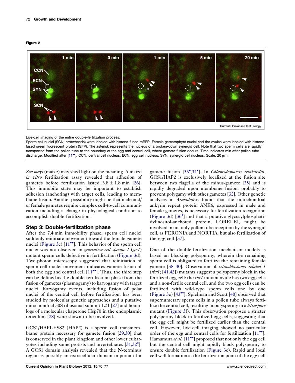
72 Growth and Development Figure2 20m ent Opinion in Plan arge rom the egg ce SYN,ynergid cll nuceus Scale,20m een two flagella of the minus-gamete [35]and is rapidly degraded upon membrane 32 probably to 3d36 Step 3:Double-fertilization phase the egg cell (37). ci (Figre c)This behavior of nuclei was no ofthedoube-mehnism On Two-photon mic oscopy sugges 40.Observation of retinoblastom fusion of gametes(plasmogamy)to karyogamy with target ferie nacland the clcan be by pemumerary sperm cells in a pollen tube always ferti- GCSI/HAPLESS2 (HAP2) ver. sed that not only the egg cel Current Opinion in Plant Biology 201.1570-7 www. edirect.com Zea mays (maize) may shed light on the meaning. A maize in vitro fertilization assay revealed that adhesion of gametes before fertilization lasted 3.8 1.8 min [26]. This immobile state may be important to establish adhesion (anchoring) with target cells, leading to membrane fusion. Another possibility might be that male and/ or female gametes require complex cell-to-cell communication including a change in physiological condition to accomplish double fertilization. Step 3: Double-fertilization phase After the 7.4 min immobility phase, sperm cell nuclei suddenly reinitiate movement toward the female gamete nuclei (Figure 3c) [11]. This behavior of the sperm cell nuclei was not observed in generative cell specific 1 (gcs1) mutant sperm cells defective in fertilization (Figure 3d). Two-photon microscopy suggested that reinitiation of sperm cell nuclei movement indicates gamete fusion of both the egg and central cell [11]. Thus, the third step can be defined as the double-fertilization phase from the fusion of gametes (plasmogamy) to karyogamy with target nuclei. Karyogamy events, including fusion of polar nuclei of the central cell before fertilization, has been studied by molecular genetic approaches and a putative mitochondrial 50S ribosomal subunit L21 [27] and homologs of a molecular chaperone Hsp70 in the endoplasmic reticulum [28] were shown to be involved. GCS1/HAPLESS2 (HAP2) is a sperm cell transmembrane protein necessary for gamete fusion [29,30] that is conserved in the plant kingdom and other lower eukaryotes including some protists and invertebrates [31,32 ]. A GCS1 domain analysis revealed that the N-terminus region is possibly an extracellular domain important for gamete fusion [33 ,34 ]. In Chlamydomonas reinhardtii, GCS1/HAP2 is exclusively localized at the fusion site between two flagella of the minus-gamete [35] and is rapidly degraded upon membrane fusion, probably to prevent polygamy with other gametes [32]. Other genetic analyses in Arabidopsis found that the mitochondrial ankyrin repeat protein ANK6, expressed in male and female gametes, is necessary for fertilization recognition (Figure 3d) [36 ] and that a putative glycosylphosphatidylinositol-anchored protein, LORELEI, might be involved in not only pollen tube reception by the synergid cell, as FERONIA and NORTIA, but also fertilization of the egg cell [37]. One of the double-fertilization mechanism models is based on blocking polyspermy, wherein the remaining sperm cell is obligated to fertilize the remaining female gamete [38–40]. Observation of retinoblastoma related 1 (rbr1; [41,42]) mutants suggest a polyspermy block in the fertilized egg cell: the rbr1 mutant ovule has two egg cells and a non-fertile central cell, and the two egg cells can be fertilized with wild-type sperm cells one by one (Figure 3e) [43]. Spielman and Scott [40] observed that supernumerary sperm cells in a pollen tube always fertilize the central cell, resulting in polyspermy in a tetraspore mutant (Figure 3f). This observation proposes a stricter polyspermy block in fertilized egg cells, suggesting that the egg cell might be fertilized earlier than the central cell. However, live-cell imaging showed no particular order of the egg and central cells for fertilization [11]. Hamamura et al. [11] proposed that not only the egg cell but the central cell might rapidly block polyspermy to ensure double fertilization (Figure 3c). Rapid and local cell wall formation at the fertilization point of the egg cell 72 Growth and Development Figure 2 -1 min 0 min 1 min 5 min 20 min CCN ECN SYN SCN Current Opinion in Plant Biology Live-cell imaging of the entire double-fertilization process. Sperm cell nuclei (SCN; arrowheads) were labeled with histone-fused mRFP. Female gametophyte nuclei and the ovules were labeled with histonefused green fluorescent protein (GFP). The asterisk represents the nucleus of a broken-down synergid cell. Note that two sperm cells are rapidly transported from the pollen tube to the boundary of the egg and central cell, where gamete fusion occurs. Time indicates min after pollen tube discharge. Modified after [11]. CCN, central cell nucleus; ECN, egg cell nucleus; SYN, synergid cell nucleus. Scale, 20 mm. Current Opinion in Plant Biology 2012, 15:70–77 www.sciencedirect.com�����������������