正在加载图片...

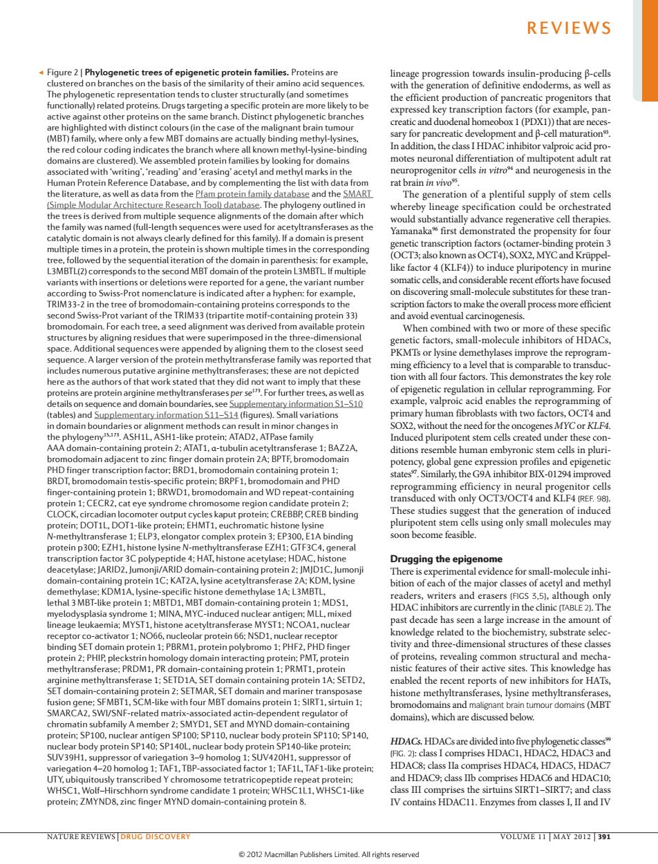
REVIEWS Figure 2|Phyloge etic tree s of epi roteinfhmilies.Protein he phy a tends to cluster st cturall (and sc the 6、 n factors (Pan homeobox 1 (PD )that arer nily.whe e only a f N MBT lly bind sI HDAC inhibitor s the ns are We as ed pr mili in motes neuron n Protein ref nce Datab a the ist with chitec Res med (full-e ially advance re erative cell the 6 ca first ys times ina tim )SOX2,MYC an ds to the nd MBT d the pr ss-Pr icated afte isco ring small-m cule st stitutes for the tran n the theTRIM33( he ov ss mor artit ot ith e of the es b imp sed combined the t .A larg the gram ansfe tive ming effic y to a level tha is comparable to transd did n in cellular re Fo cna the 89 mer methodsca h vithout the need for t nesMYCor KLF4 TAD nduced pluri potent stem s crea 2 bal g files and epigeneti 01294 npro nd WD with onl REF 98 CLOCK ioaoinduce p30 m feasible e:AF om ety ein ce for small-molecule inhi the ma anc ethy thylas rs are cu n the clinic MINA duced nu MYST1:NCOA e amount o rotein 6 d three-dimensional structur s of these cla ro Z:PH P.ple logy ain int ing onta ETMA enabled the recent reports of new inhibitors for HATs h four MB s protein 1: s and mali domains),which are discussed below. YD and MY 140 SP140 40 HDACs.HDACsare dividedin o five ph lear body ike prote 2):class I comprises HD HD TAFI-ike protein DAC 1og 1:d AF1.TBP-as factor 1 and HDAC9;class IIb s HDAC and HDACIO - ate WHSC1L1.WHSC1-like T7ianddas NATURE REVIEWSIDRUG DISCOVERY VOLUME 11 MAY 2012 391▶ Figure 2 | Phylogenetic trees of epigenetic protein families. Proteins are clustered on branches on the basis of the similarity of their amino acid sequences. The phylogenetic representation tends to cluster structurally (and sometimes functionally) related proteins. Drugs targeting a specific protein are more likely to be active against other proteins on the same branch. Distinct phylogenetic branches are highlighted with distinct colours (in the case of the malignant brain tumour (MBT) family, where only a few MBT domains are actually binding methyl-lysines, the red colour coding indicates the branch where all known methyl-lysine-binding domains are clustered). We assembled protein families by looking for domains associated with ‘writing’, ‘reading’ and ‘erasing’ acetyl and methyl marks in the Human Protein Reference Database, and by complementing the list with data from the literature, as well as data from the Pfam protein family database and the SMART (Simple Modular Architecture Research Tool) database. The phylogeny outlined in the trees is derived from multiple sequence alignments of the domain after which the family was named (full-length sequences were used for acetyltransferases as the catalytic domain is not always clearly defined for this family). If a domain is present multiple times in a protein, the protein is shown multiple times in the corresponding tree, followed by the sequential iteration of the domain in parenthesis: for example, L3MBTL(2) corresponds to the second MBT domain of the protein L3MBTL. If multiple variants with insertions or deletions were reported for a gene, the variant number according to Swiss-Prot nomenclature is indicated after a hyphen: for example, TRIM33-2 in the tree of bromodomain-containing proteins corresponds to the second Swiss-Prot variant of the TRIM33 (tripartite motif-containing protein 33) bromodomain. For each tree, a seed alignment was derived from available protein structures by aligning residues that were superimposed in the three-dimensional space. Additional sequences were appended by aligning them to the closest seed sequence. A larger version of the protein methyltransferase family was reported that includes numerous putative arginine methyltransferases; these are not depicted here as the authors of that work stated that they did not want to imply that these proteins are protein arginine methyltransferases per se173. For further trees, as well as details on sequence and domain boundaries, see Supplementary information S1–S10 (tables) and Supplementary information S11–S14 (figures). Small variations in domain boundaries or alignment methods can result in minor changes in the phylogeny15,173. ASH1L, ASH1-like protein; ATAD2, ATPase family AAA domain-containing protein 2; ATAT1, α-tubulin acetyltransferase 1; BAZ2A, bromodomain adjacent to zinc finger domain protein 2A; BPTF, bromodomain PHD finger transcription factor; BRD1, bromodomain containing protein 1; BRDT, bromodomain testis-specific protein; BRPF1, bromodomain and PHD finger-containing protein 1; BRWD1, bromodomain and WD repeat-containing protein 1; CECR2, cat eye syndrome chromosome region candidate protein 2; CLOCK, circadian locomoter output cycles kaput protein; CREBBP, CREB binding protein; DOT1L, DOT1-like protein; EHMT1, euchromatic histone lysine N-methyltransferase 1; ELP3, elongator complex protein 3; EP300, E1A binding protein p300; EZH1, histone lysine N-methyltransferase EZH1; GTF3C4, general transcription factor 3C polypeptide 4; HAT, histone acetylase; HDAC, histone deacetylase; JARID2, Jumonji/ARID domain-containing protein 2; JMJD1C, Jumonji domain-containing protein 1C; KAT2A, lysine acetyltransferase 2A; KDM, lysine demethylase; KDM1A, lysine-specific histone demethylase 1A; L3MBTL, lethal 3 MBT-like protein 1; MBTD1, MBT domain-containing protein 1; MDS1, myelodysplasia syndrome 1; MINA, MYC-induced nuclear antigen; MLL, mixed lineage leukaemia; MYST1, histone acetyltransferase MYST1; NCOA1, nuclear receptor co-activator 1; NO66, nucleolar protein 66; NSD1, nuclear receptor binding SET domain protein 1; PBRM1, protein polybromo 1; PHF2, PHD finger protein 2; PHIP, pleckstrin homology domain interacting protein; PMT, protein methyltransferase; PRDM1, PR domain-containing protein 1; PRMT1, protein arginine methyltransferase 1; SETD1A, SET domain containing protein 1A; SETD2, SET domain-containing protein 2; SETMAR, SET domain and mariner transposase fusion gene; SFMBT1, SCM-like with four MBT domains protein 1; SIRT1, sirtuin 1; SMARCA2, SWI/SNF-related matrix-associated actin-dependent regulator of chromatin subfamily A member 2; SMYD1, SET and MYND domain-containing protein; SP100, nuclear antigen SP100; SP110, nuclear body protein SP110; SP140, nuclear body protein SP140; SP140L, nuclear body protein SP140‑like protein; SUV39H1, suppressor of variegation 3–9 homolog 1; SUV420H1, suppressor of variegation 4–20 homolog 1; TAF1, TBP-associated factor 1; TAF1L, TAF1‑like protein; UTY, ubiquitously transcribed Y chromosome tetratricopeptide repeat protein; WHSC1, Wolf–Hirschhorn syndrome candidate 1 protein; WHSC1L1, WHSC1-like protein; ZMYND8, zinc finger MYND domain-containing protein 8. lineage progression towards insulin-producing β-cells with the generation of definitive endoderms, as well as the efficient production of pancreatic progenitors that expressed key transcription factors (for example, pancreatic and duodenal homeobox 1 (PDX1)) that are necessary for pancreatic development and β-cell maturation93. In addition, the class I HDAC inhibitor valproic acid promotes neuronal differentiation of multipotent adult rat neuroprogenitor cells in vitro94 and neurogenesis in the rat brain in vivo95. The generation of a plentiful supply of stem cells whereby lineage specification could be orchestrated would substantially advance regenerative cell therapies. Yamanaka96 first demonstrated the propensity for four genetic transcription factors (octamer-binding protein 3 (OCT3; also known as OCT4), SOX2, MYC and Krüppellike factor 4 (KLF4)) to induce pluripotency in murine somatic cells, and considerable recent efforts have focused on discovering small-molecule substitutes for these transcription factors to make the overall process more efficient and avoid eventual carcinogenesis. When combined with two or more of these specific genetic factors, small-molecule inhibitors of HDACs, PKMTs or lysine demethylases improve the reprogramming efficiency to a level that is comparable to transduction with all four factors. This demonstrates the key role of epigenetic regulation in cellular reprogramming. For example, valproic acid enables the reprogramming of primary human fibroblasts with two factors, OCT4 and SOX2, without the need for the oncogenes MYC or KLF4. Induced pluripotent stem cells created under these conditions resemble human embyronic stem cells in pluripotency, global gene expression profiles and epigenetic states97. Similarly, the G9A inhibitor BIX-01294 improved reprogramming efficiency in neural progenitor cells transduced with only OCT3/OCT4 and KLF4 (REF. 98). These studies suggest that the generation of induced pluripotent stem cells using only small molecules may soon become feasible. Drugging the epigenome There is experimental evidence for small-molecule inhibition of each of the major classes of acetyl and methyl readers, writers and erasers (FIGS 3,5), although only HDAC inhibitors are currently in the clinic (TABLE 2). The past decade has seen a large increase in the amount of knowledge related to the biochemistry, substrate selectivity and three-dimensional structures of these classes of proteins, revealing common structural and mechanistic features of their active sites. This knowledge has enabled the recent reports of new inhibitors for HATs, histone methyltransferases, lysine methyltransferases, bromodomains and malignant brain tumour domains (MBT domains), which are discussed below. HDACs. HDACs are divided into five phylogenetic classes99 (FIG. 2): class I comprises HDAC1, HDAC2, HDAC3 and HDAC8; class IIa comprises HDAC4, HDAC5, HDAC7 and HDAC9; class IIb comprises HDAC6 and HDAC10; class III comprises the sirtuins SIRT1–SIRT7; and class IV contains HDAC11. Enzymes from classes I, II and IV REVIEWS NATURE REVIEWS | DRUG DISCOVERY VOLUME 11 | MAY 2012 | 391 © 2012 Macmillan Publishers Limited. All rights reserved