正在加载图片...
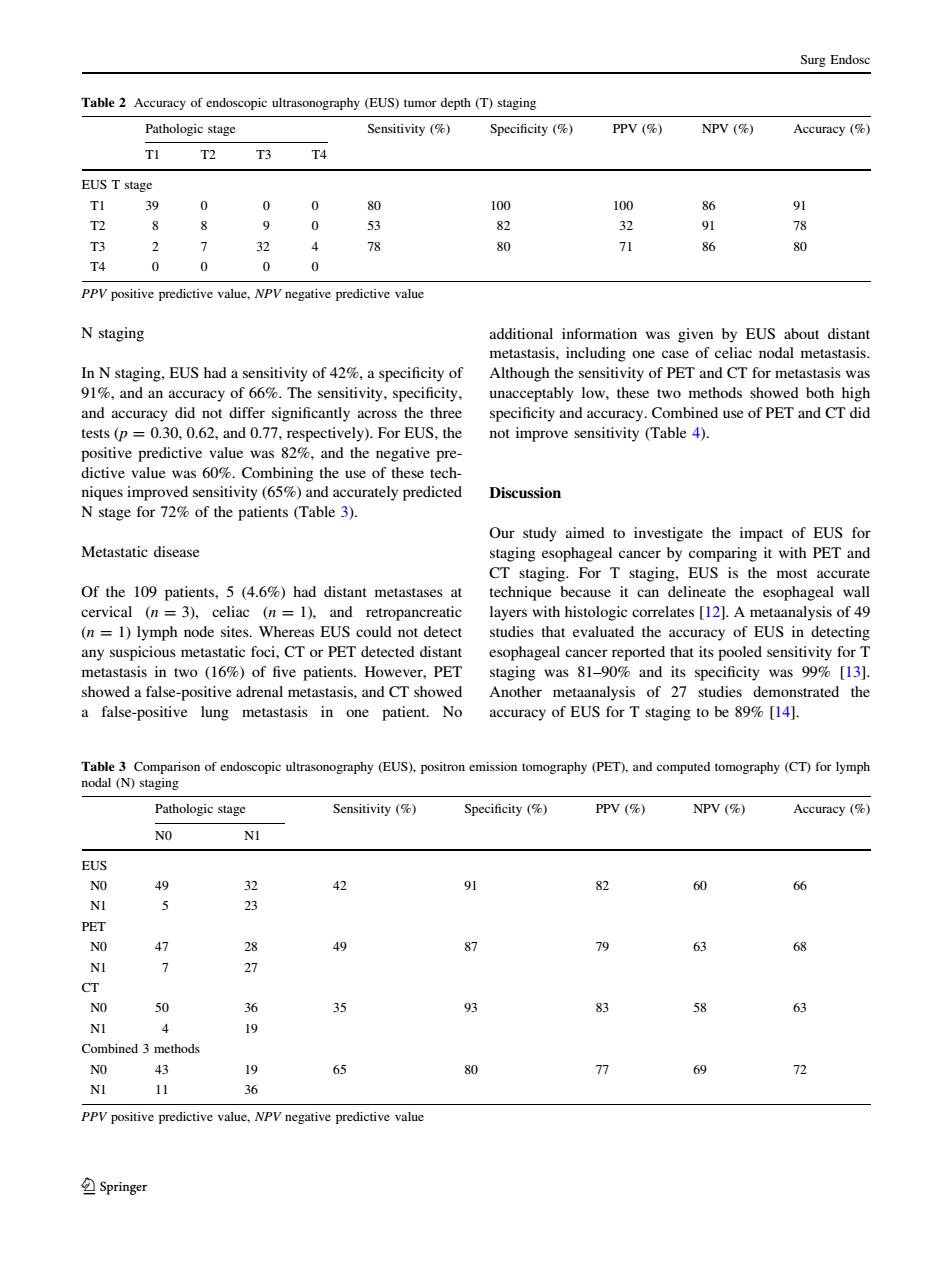
Surg Endose Table2 Accuracy of endoscopic ultrasonography(EUS)tumor depth(T)staging Pathologic stage Sensitivity(候) Specificity (% PPV (% NPV ( Accuracy (% T3 T4 EUS T stage 30 0 0 1 86 91 T2 8 8 9 0 5 82 78 T3 2 32 4 8 80 71 86 80 T4 0 0 0 0 N staging additional information was given by EUS about distan metastasis,including one case of celiac nodal metastasis In N staging.EUS had a sensitivity of 42,a specificity of Although the sensitivity of PET and CT for metastasis was 1and an accuracy of%The sensitivity,specificity. unacceptably low,these edictive value was 82 and the dictive value was 60%.Combining the use of these tech niques improved sensitivity (65%)and accurately predicted Discussion N stage for 72%of the patients (Table 3). Metastatic disease Of the 109 patients.5 (4.6)had distant metastases at ageal wal cervical (n=3)celiac (n=1)and retropancreatic layers with histologie correlates [12.A metaanalysis of 49 (n=1)lymph node sites.Whereas EUS could not detect studies that evaluated the accuracy of EUS in detecting any suspicious metastatic foci,CT or PET detected distant esophageal cancer reported that its pooled sensitivity for taging was 81- false in one uracy o 899%14 strated the opic ul raphy(EUS). pos graphy (PET) and Pathologic stage PPV ( NPV ( Accuracy ( NO NI EUS 3 0 66 47 28 40 NI 7 27 CT NO 50 36 93 58 63 NI 19 bined 3 methods 19 65 69 72 30 PPV positive predictive value.NPV negative predictive value SpringerN staging In N staging, EUS had a sensitivity of 42%, a specificity of 91%, and an accuracy of 66%. The sensitivity, specificity, and accuracy did not differ significantly across the three tests (p = 0.30, 0.62, and 0.77, respectively). For EUS, the positive predictive value was 82%, and the negative predictive value was 60%. Combining the use of these techniques improved sensitivity (65%) and accurately predicted N stage for 72% of the patients (Table 3). Metastatic disease Of the 109 patients, 5 (4.6%) had distant metastases at cervical (n = 3), celiac (n = 1), and retropancreatic (n = 1) lymph node sites. Whereas EUS could not detect any suspicious metastatic foci, CT or PET detected distant metastasis in two (16%) of five patients. However, PET showed a false-positive adrenal metastasis, and CT showed a false-positive lung metastasis in one patient. No additional information was given by EUS about distant metastasis, including one case of celiac nodal metastasis. Although the sensitivity of PET and CT for metastasis was unacceptably low, these two methods showed both high specificity and accuracy. Combined use of PET and CT did not improve sensitivity (Table 4). Discussion Our study aimed to investigate the impact of EUS for staging esophageal cancer by comparing it with PET and CT staging. For T staging, EUS is the most accurate technique because it can delineate the esophageal wall layers with histologic correlates [12]. A metaanalysis of 49 studies that evaluated the accuracy of EUS in detecting esophageal cancer reported that its pooled sensitivity for T staging was 81–90% and its specificity was 99% [13]. Another metaanalysis of 27 studies demonstrated the accuracy of EUS for T staging to be 89% [14]. Table 2 Accuracy of endoscopic ultrasonography (EUS) tumor depth (T) staging Pathologic stage Sensitivity (%) Specificity (%) PPV (%) NPV (%) Accuracy (%) T1 T2 T3 T4 EUS T stage T1 39 0 0 0 80 100 100 86 91 T2 8 8 9 0 53 82 32 91 78 T3 2 7 32 4 78 80 71 86 80 T4 0 0 0 0 PPV positive predictive value, NPV negative predictive value Table 3 Comparison of endoscopic ultrasonography (EUS), positron emission tomography (PET), and computed tomography (CT) for lymph nodal (N) staging Pathologic stage Sensitivity (%) Specificity (%) PPV (%) NPV (%) Accuracy (%) N0 N1 EUS N0 49 32 42 91 82 60 66 N1 5 23 PET N0 47 28 49 87 79 63 68 N1 7 27 CT N0 50 36 35 93 83 58 63 N1 4 19 Combined 3 methods N0 43 19 65 80 77 69 72 N1 11 36 PPV positive predictive value, NPV negative predictive value Surg Endosc 123