正在加载图片...
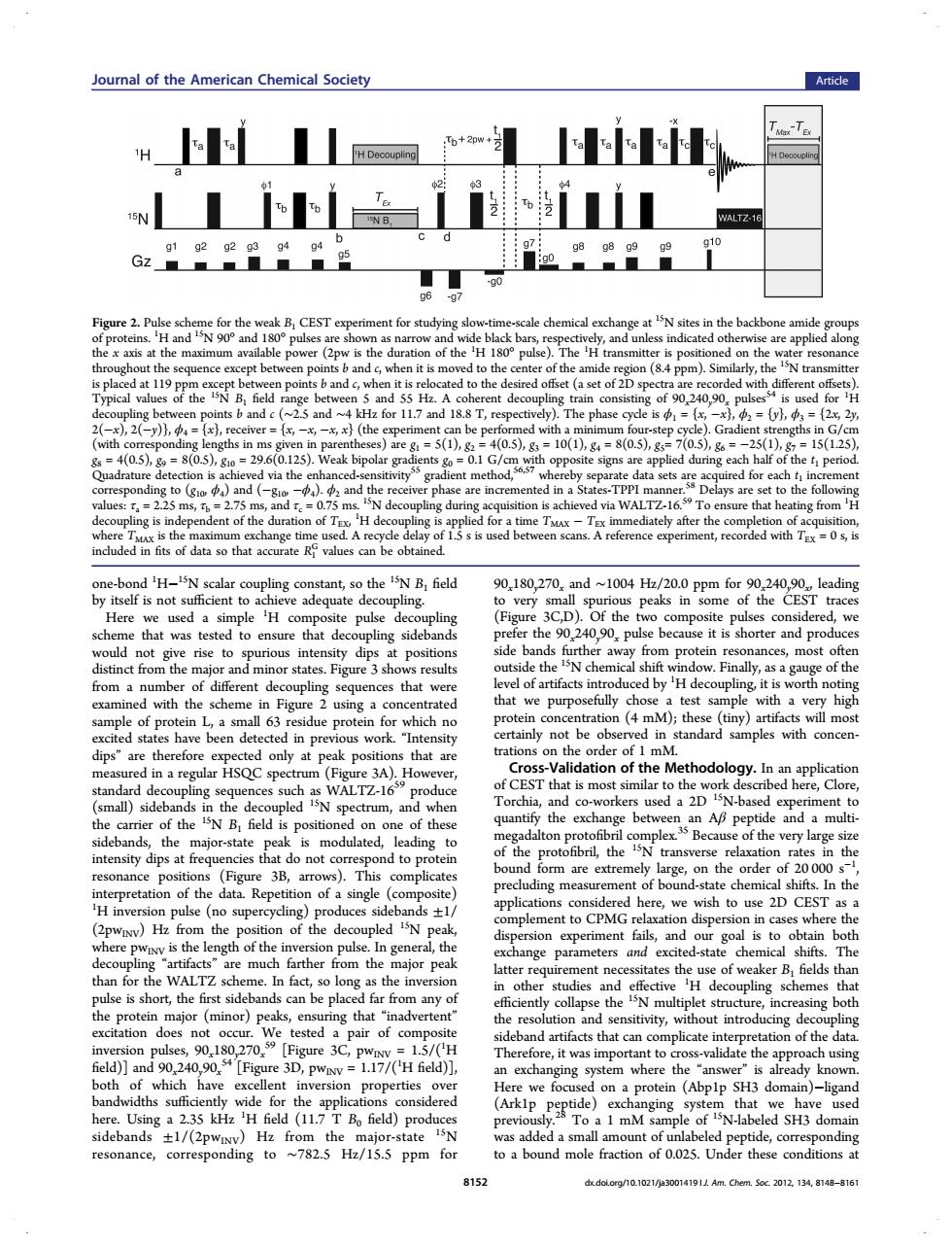
oural of the American Chemical Society Article H网 H LLiraLil WALTZ- k B.CEST N90°and18 pu are appl the e)The H d at the (a set of2Ds ded with击d a117 1188T s,= hs in (0s) to and d th of t tely after t ecorded with in fts of data so that a ate H PN scalar o the B.field 90,180,270,and~1004Hh/20.0 Ppm for90,240,90。l1 eading idered horter and pr A f th purp chos est sampl with of p 63 ich n on(年m ny) a regula rum 3A).Hov of C NB field is positione AB tide and a multi carier of the thes f the very large sity dips at fr order 000 e posof th here.we wish to use 2D CEST SuD side nple h eg ange eters s and exc ed-stat che Th rthe oupling sc s tha t the and effe the he protein najor (minor)peak ng tha tion and 00102 tifacts that can complicate i a d)an 024090, re 3D,pwv=1.17/('H field)] ystem where th is a dy k Bo field) dom 191 one-bond 1 H−15N scalar coupling constant, so the 15N B1 field by itself is not sufficient to achieve adequate decoupling. Here we used a simple 1 H composite pulse decoupling scheme that was tested to ensure that decoupling sidebands would not give rise to spurious intensity dips at positions distinct from the major and minor states. Figure 3 shows results from a number of different decoupling sequences that were examined with the scheme in Figure 2 using a concentrated sample of protein L, a small 63 residue protein for which no excited states have been detected in previous work. “Intensity dips” are therefore expected only at peak positions that are measured in a regular HSQC spectrum (Figure 3A). However, standard decoupling sequences such as WALTZ-1659 produce (small) sidebands in the decoupled 15N spectrum, and when the carrier of the 15N B1 field is positioned on one of these sidebands, the major-state peak is modulated, leading to intensity dips at frequencies that do not correspond to protein resonance positions (Figure 3B, arrows). This complicates interpretation of the data. Repetition of a single (composite) 1 H inversion pulse (no supercycling) produces sidebands ±1/ (2pwINV) Hz from the position of the decoupled 15N peak, where pwINV is the length of the inversion pulse. In general, the decoupling “artifacts” are much farther from the major peak than for the WALTZ scheme. In fact, so long as the inversion pulse is short, the first sidebands can be placed far from any of the protein major (minor) peaks, ensuring that “inadvertent” excitation does not occur. We tested a pair of composite inversion pulses, 90x180y270x 59 [Figure 3C, pwINV = 1.5/(1 H field)] and 90x240y90x 54 [Figure 3D, pwINV = 1.17/(1 H field)], both of which have excellent inversion properties over bandwidths sufficiently wide for the applications considered here. Using a 2.35 kHz 1 H field (11.7 T B0 field) produces sidebands ±1/(2pwINV) Hz from the major-state 15N resonance, corresponding to ∼782.5 Hz/15.5 ppm for 90x180y270x and ∼1004 Hz/20.0 ppm for 90x240y90x, leading to very small spurious peaks in some of the CEST traces (Figure 3C,D). Of the two composite pulses considered, we prefer the 90x240y90x pulse because it is shorter and produces side bands further away from protein resonances, most often outside the 15N chemical shift window. Finally, as a gauge of the level of artifacts introduced by 1 H decoupling, it is worth noting that we purposefully chose a test sample with a very high protein concentration (4 mM); these (tiny) artifacts will most certainly not be observed in standard samples with concentrations on the order of 1 mM. Cross-Validation of the Methodology. In an application of CEST that is most similar to the work described here, Clore, Torchia, and co-workers used a 2D 15N-based experiment to quantify the exchange between an Aβ peptide and a multimegadalton protofibril complex.35 Because of the very large size of the protofibril, the 15N transverse relaxation rates in the bound form are extremely large, on the order of 20 000 s−1 , precluding measurement of bound-state chemical shifts. In the applications considered here, we wish to use 2D CEST as a complement to CPMG relaxation dispersion in cases where the dispersion experiment fails, and our goal is to obtain both exchange parameters and excited-state chemical shifts. The latter requirement necessitates the use of weaker B1 fields than in other studies and effective 1 H decoupling schemes that efficiently collapse the 15N multiplet structure, increasing both the resolution and sensitivity, without introducing decoupling sideband artifacts that can complicate interpretation of the data. Therefore, it was important to cross-validate the approach using an exchanging system where the “answer” is already known. Here we focused on a protein (Abp1p SH3 domain)−ligand (Ark1p peptide) exchanging system that we have used previously.28 To a 1 mM sample of 15N-labeled SH3 domain was added a small amount of unlabeled peptide, corresponding to a bound mole fraction of 0.025. Under these conditions at Figure 2. Pulse scheme for the weak B1 CEST experiment for studying slow-time-scale chemical exchange at 15N sites in the backbone amide groups of proteins. 1 H and 15N 90° and 180° pulses are shown as narrow and wide black bars, respectively, and unless indicated otherwise are applied along the x axis at the maximum available power (2pw is the duration of the 1 H 180° pulse). The 1 H transmitter is positioned on the water resonance throughout the sequence except between points b and c, when it is moved to the center of the amide region (8.4 ppm). Similarly, the 15N transmitter is placed at 119 ppm except between points b and c, when it is relocated to the desired offset (a set of 2D spectra are recorded with different offsets). Typical values of the 15N B1 field range between 5 and 55 Hz. A coherent decoupling train consisting of 90x240y90x pulses54 is used for 1 H decoupling between points b and c (∼2.5 and ∼4 kHz for 11.7 and 18.8 T, respectively). The phase cycle is ϕ1 = {x, −x}, ϕ2 = {y}, ϕ3 = {2x, 2y, 2(−x), 2(−y)}, ϕ4 = {x}, receiver = {x, −x, −x, x} (the experiment can be performed with a minimum four-step cycle). Gradient strengths in G/cm (with corresponding lengths in ms given in parentheses) are g1 = 5(1), g2 = 4(0.5), g3 = 10(1), g4 = 8(0.5), g5= 7(0.5), g6 = −25(1), g7 = 15(1.25), g8 = 4(0.5), g9 = 8(0.5), g10 = 29.6(0.125). Weak bipolar gradients g0 = 0.1 G/cm with opposite signs are applied during each half of the t1 period. Quadrature detection is achieved via the enhanced-sensitivity55 gradient method,56,57 whereby separate data sets are acquired for each t1 increment corresponding to (g10, ϕ4) and (−g10, −ϕ4). ϕ2 and the receiver phase are incremented in a States-TPPI manner.58 Delays are set to the following values: τa = 2.25 ms, τb = 2.75 ms, and τc = 0.75 ms. 15N decoupling during acquisition is achieved via WALTZ-16.59 To ensure that heating from 1 H decoupling is independent of the duration of TEX, 1 H decoupling is applied for a time TMAX − TEX immediately after the completion of acquisition, where TMAX is the maximum exchange time used. A recycle delay of 1.5 s is used between scans. A reference experiment, recorded with TEX = 0 s, is included in fits of data so that accurate R1 G values can be obtained. Journal of the American Chemical Society Article 8152 dx.doi.org/10.1021/ja3001419 | J. Am. Chem. Soc. 2012, 134, 8148−8161