正在加载图片...
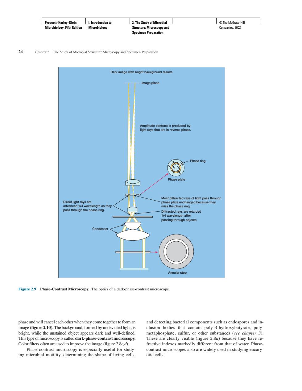
eh” eeaeaeoee Phase plate the phasea ass the Figure2 Phase-Contrast Microscopy.The optics ofa dark-phase-contrast microscope. phase and will can eleach other when they come together to form an and detecting bacterial components such as endospores and in m and well-detined These are clearly visible (figure2.8d)because they havere Phas otrast microscon y is especially useful for study- ntras microscopes also are widely used in studying eucary ing microbial motility,determining the shape of living cells. otic cells. Prescott−Harley−Klein: Microbiology, Fifth Edition I. Introduction to Microbiology 2. The Study of Microbial Structure: Microscopy and Specimen Preparation © The McGraw−Hill Companies, 2002 phase and will cancel each other when they come together to form an image (figure 2.10). The background, formed by undeviated light, is bright, while the unstained object appears dark and well-defined. This type of microscopy is called dark-phase-contrast microscopy. Color filters often are used to improve the image (figure 2.8c,d). Phase-contrast microscopy is especially useful for studying microbial motility, determining the shape of living cells, and detecting bacterial components such as endospores and inclusion bodies that contain poly- -hydroxybutyrate, polymetaphosphate, sulfur, or other substances (see chapter 3). These are clearly visible (figure 2.8d) because they have refractive indexes markedly different from that of water. Phasecontrast microscopes also are widely used in studying eucaryotic cells. 24 Chapter 2 The Study of Microbial Structure: Microscopy and Specimen Preparation Dark image with bright background results Image plane Amplitude contrast is produced by light rays that are in reverse phase. Phase ring Phase plate Most diffracted rays of light pass through phase plate unchanged because they miss the phase ring. Diffracted rays are retarded 1/4 wavelength after passing through objects. Annular stop Condenser Direct light rays are advanced 1/4 wavelength as they pass through the phase ring. Figure 2.9 Phase-Contrast Microscopy. The optics of a dark-phase-contrast microscope