正在加载图片...
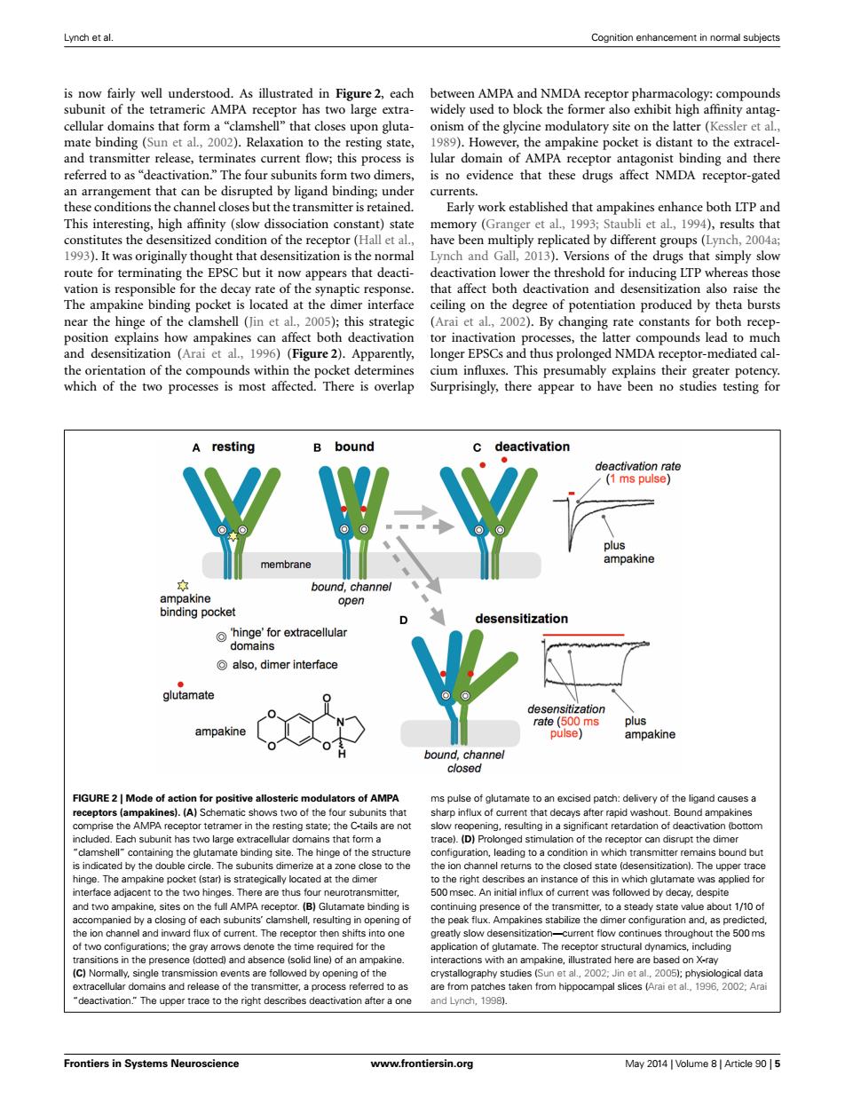
Lynch et al. Cognition enhancement in normal subjects is now fairly well understood.As illustrated in Figure 2,each between AMPA and NMDA receptor pharmacology:compounds subunit of the tetrameric AMPA receptor has two large extra- widely used to block the former also exhibit high affinity antag- cellular domains that form a"clamshell"that closes upon gluta- onism of the glycine modulatory site on the latter(Kessler et al., mate binding (Sun et al.,2002).Relaxation to the resting state, 1989).However,the ampakine pocket is distant to the extracel- and transmitter release,terminates current flow;this process is lular domain of AMPA receptor antagonist binding and there referred to as"deactivation."The four subunits form two dimers,is no evidence that these drugs affect NMDA receptor-gated an arrangement that can be disrupted by ligand binding;under currents. these conditions the channel closes but the transmitter is retained. Early work established that ampakines enhance both LIP and This interesting,high affinity (slow dissociation constant)state memory (Granger et al.,1993;Staubli et al.,1994),results that constitutes the desensitized condition of the receptor (Hall et al., have been multiply replicated by different groups(Lynch,2004a; 1993).It was originally thought that desensitization is the normal Lynch and Gall,2013).Versions of the drugs that simply slow route for terminating the EPSC but it now appears that deacti-deactivation lower the threshold for inducing LTP whereas those vation is responsible for the decay rate of the synaptic response. that affect both deactivation and desensitization also raise the The ampakine binding pocket is located at the dimer interface ceiling on the degree of potentiation produced by theta bursts near the hinge of the clamshell(Jin et al,2005);this strategic (Arai et al,2002).By changing rate constants for both recep- position explains how ampakines can affect both deactivation tor inactivation processes,the latter compounds lead to much and desensitization (Arai et al.,1996)(Figure 2).Apparently, longer EPSCs and thus prolonged NMDA receptor-mediated cal- the orientation of the compounds within the pocket determines cium influxes.This presumably explains their greater potency. which of the two processes is most affected.There is overlap Surprisingly,there appear to have been no studies testing for A resting bound c deactivation deactivation rate (1 ms pulse) plus membrane ampakine 的 bound,channel ampakine open binding pocket desensitization hinge'for extracellular domains also,dimer interface glutamate desensitization rate(500 ms plus ampakine pulse) ampakine bound,channel closed FIGURE 2 Mode of action for positive allosteric modulators of AMPA ms pulse of glutamate to an excised patch:delivery of the ligand causes a receptors (ampakines).(A)Schematic shows two of the four subunits that sharp influx of current that decays after rapid washout.Bound ampakines comprise the AMPA receptor tetramer in the resting state;the C-tails are not slow reopening,resulting in a significant retardation of deactivation (bottom included.Each subunit has two large extracellular domains that form a trace).(D)Prolonged stimulation of the receptor can disrupt the dimer "clamshell"containing the glutamate binding site.The hinge of the structure configuration,leading to a condition in which transmitter remains bound but is indicated by the double circle.The subunits dimerize at a zone close to the the ion channel returns to the closed state (desensitization).The upper trace hinge.The ampakine pocket (star)is strategically located at the dimer to the right describes an instance of this in which glutamate was applied for interface adjacent to the two hinges.There are thus four neurotransmitter, 500 msec.An initial influx of current was followed by decay,despite and two ampakine,sites on the full AMPA receptor.(B)Glutamate binding is continuing presence of the transmitter,to a steady state value about 1/10 of accompanied by a closing of each subunits'clamshell,resulting in opening of the peak flux.Ampakines stabilize the dimer configuration and,as predicted, the ion channel and inward flux of current.The receptor then shifts into one greatly slow desensitization-current flow continues throughout the 500 ms of two configurations;the gray arrows denote the time required for the application of glutamate.The receptor structural dynamics,including transitions in the presence (dotted)and absence (solid line)of an ampakine. interactions with an ampakine,illustrated here are based on X-ray (C)Normally,single transmission events are followed by opening of the crystallography studies (Sun et al.,2002:Jin et al.,2005);physiological data extracellular domains and release of the transmitter,a process referred to as are from patches taken from hippocampal slices (Arai et al..1996.2002:Arai 'deactivation."The upper trace to the right describes deactivation after a one and Lynch,1998). Frontiers in Systems Neuroscience www.frontiersin.org May 2014 Volume 8 Article 905Lynch et al. Cognition enhancement in normal subjects is now fairly well understood. As illustrated in Figure 2, each subunit of the tetrameric AMPA receptor has two large extracellular domains that form a “clamshell” that closes upon glutamate binding (Sun et al., 2002). Relaxation to the resting state, and transmitter release, terminates current flow; this process is referred to as “deactivation.” The four subunits form two dimers, an arrangement that can be disrupted by ligand binding; under these conditions the channel closes but the transmitter is retained. This interesting, high affinity (slow dissociation constant) state constitutes the desensitized condition of the receptor (Hall et al., 1993). It was originally thought that desensitization is the normal route for terminating the EPSC but it now appears that deactivation is responsible for the decay rate of the synaptic response. The ampakine binding pocket is located at the dimer interface near the hinge of the clamshell (Jin et al., 2005); this strategic position explains how ampakines can affect both deactivation and desensitization (Arai et al., 1996) (Figure 2). Apparently, the orientation of the compounds within the pocket determines which of the two processes is most affected. There is overlap between AMPA and NMDA receptor pharmacology: compounds widely used to block the former also exhibit high affinity antagonism of the glycine modulatory site on the latter (Kessler et al., 1989). However, the ampakine pocket is distant to the extracellular domain of AMPA receptor antagonist binding and there is no evidence that these drugs affect NMDA receptor-gated currents. Early work established that ampakines enhance both LTP and memory (Granger et al., 1993; Staubli et al., 1994), results that have been multiply replicated by different groups (Lynch, 2004a; Lynch and Gall, 2013). Versions of the drugs that simply slow deactivation lower the threshold for inducing LTP whereas those that affect both deactivation and desensitization also raise the ceiling on the degree of potentiation produced by theta bursts (Arai et al., 2002). By changing rate constants for both receptor inactivation processes, the latter compounds lead to much longer EPSCs and thus prolonged NMDA receptor-mediated calcium influxes. This presumably explains their greater potency. Surprisingly, there appear to have been no studies testing for FIGURE 2 | Mode of action for positive allosteric modulators of AMPA receptors (ampakines). (A) Schematic shows two of the four subunits that comprise the AMPA receptor tetramer in the resting state; the C-tails are not included. Each subunit has two large extracellular domains that form a “clamshell” containing the glutamate binding site. The hinge of the structure is indicated by the double circle. The subunits dimerize at a zone close to the hinge. The ampakine pocket (star) is strategically located at the dimer interface adjacent to the two hinges. There are thus four neurotransmitter, and two ampakine, sites on the full AMPA receptor. (B) Glutamate binding is accompanied by a closing of each subunits’ clamshell, resulting in opening of the ion channel and inward flux of current. The receptor then shifts into one of two configurations; the gray arrows denote the time required for the transitions in the presence (dotted) and absence (solid line) of an ampakine. (C) Normally, single transmission events are followed by opening of the extracellular domains and release of the transmitter, a process referred to as “deactivation.” The upper trace to the right describes deactivation after a one ms pulse of glutamate to an excised patch: delivery of the ligand causes a sharp influx of current that decays after rapid washout. Bound ampakines slow reopening, resulting in a significant retardation of deactivation (bottom trace). (D) Prolonged stimulation of the receptor can disrupt the dimer configuration, leading to a condition in which transmitter remains bound but the ion channel returns to the closed state (desensitization). The upper trace to the right describes an instance of this in which glutamate was applied for 500 msec. An initial influx of current was followed by decay, despite continuing presence of the transmitter, to a steady state value about 1/10 of the peak flux. Ampakines stabilize the dimer configuration and, as predicted, greatly slow desensitization—current flow continues throughout the 500 ms application of glutamate. The receptor structural dynamics, including interactions with an ampakine, illustrated here are based on X-ray crystallography studies (Sun et al., 2002; Jin et al., 2005); physiological data are from patches taken from hippocampal slices (Arai et al., 1996, 2002; Arai and Lynch, 1998). Frontiers in Systems Neuroscience www.frontiersin.org May 2014 | Volume 8 | Article 90 | 5