正在加载图片...
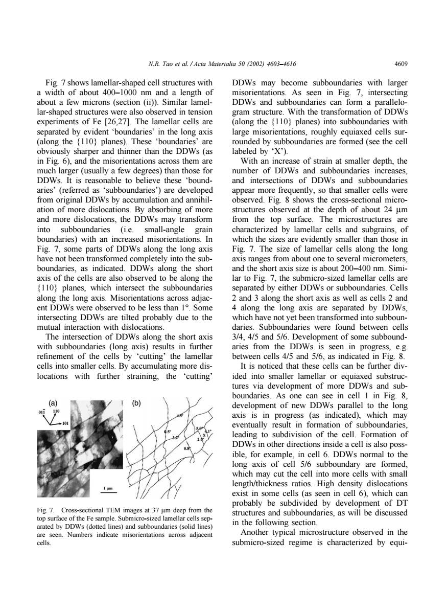
N.R.Tao et al.Acta Materialia 50 (2002)4603-4616 4609 Fig.7 shows lamellar-shaped cell structures with DDWs may become subboundaries with larger a width of about 400-1000 nm and a length of misorientations.As seen in Fig.7,intersecting about a few microns (section (ii)).Similar lamel- DDWs and subboundaries can form a parallelo- lar-shaped structures were also observed in tension gram structure.With the transformation of DDWs experiments of Fe [26,27].The lamellar cells are (along the {110)planes)into subboundaries with separated by evident boundaries'in the long axis large misorientations,roughly equiaxed cells sur- (along the (110)planes).These 'boundaries'are rounded by subboundaries are formed (see the cell obviously sharper and thinner than the DDWs(as labeled by 'X'). in Fig.6),and the misorientations across them are With an increase of strain at smaller depth,the much larger(usually a few degrees)than those for number of DDWs and subboundaries increases, DDWs.It is reasonable to believe these 'bound- and intersections of DDWs and subboundaries aries'(referred as 'subboundaries')are developed appear more frequently,so that smaller cells were from original DDWs by accumulation and annihil- observed.Fig.8 shows the cross-sectional micro- ation of more dislocations.By absorbing of more structures observed at the depth of about 24 um and more dislocations,the DDWs may transform from the top surface.The microstructures are into subboundaries (i.e.small-angle grain characterized by lamellar cells and subgrains,of boundaries)with an increased misorientations.In which the sizes are evidently smaller than those in Fig.7,some parts of DDWs along the long axis Fig.7.The size of lamellar cells along the long have not been transformed completely into the sub- axis ranges from about one to several micrometers, boundaries,as indicated.DDWs along the short and the short axis size is about 200-400 nm.Simi- axis of the cells are also observed to be along the lar to Fig.7,the submicro-sized lamellar cells are (110)planes,which intersect the subboundaries separated by either DDWs or subboundaries.Cells along the long axis.Misorientations across adjac- 2 and 3 along the short axis as well as cells 2 and ent DDWs were observed to be less than 1.Some 4 along the long axis are separated by DDWs, intersecting DDWs are tilted probably due to the which have not yet been transformed into subboun- mutual interaction with dislocations. daries.Subboundaries were found between cells The intersection of DDWs along the short axis 3/4,4/5 and 5/6.Development of some subbound- with subboundaries (long axis)results in further aries from the DDWs is seen in progress,e.g. refinement of the cells by 'cutting'the lamellar between cells 4/5 and 5/6,as indicated in Fig.8. cells into smaller cells.By accumulating more dis- It is noticed that these cells can be further div- locations with further straining,the cutting' ided into smaller lamellar or equiaxed substruc- tures via development of more DDWs and sub- boundaries.As one can see in cell 1 in Fig.8, b development of new DDWs parallel to the long axis is in progress (as indicated),which may eventually result in formation of subboundaries, leading to subdivision of the cell.Formation of DDWs in other directions inside a cell is also poss- ible,for example,in cell 6.DDWs normal to the long axis of cell 5/6 subboundary are formed, which may cut the cell into more cells with small length/thickness ratios.High density dislocations 1画 exist in some cells (as seen in cell 6),which can probably be subdivided by development of DT Fig.7.Cross-sectional TEM images at 37 um deep from the structures and subboundaries,as will be discussed top surface of the Fe sample.Submicro-sized lamellar cells sep- arated by DDWs (dotted lines)and subboundaries (solid lines) in the following section. are seen.Numbers indicate misorientations across adjacent Another typical microstructure observed in the cells. submicro-sized regime is characterized by equi-N.R. Tao et al. / Acta Materialia 50 (2002) 4603–4616 4609 Fig. 7 shows lamellar-shaped cell structures with a width of about 400–1000 nm and a length of about a few microns (section (ii)). Similar lamellar-shaped structures were also observed in tension experiments of Fe [26,27]. The lamellar cells are separated by evident ‘boundaries’ in the long axis (along the {110} planes). These ‘boundaries’ are obviously sharper and thinner than the DDWs (as in Fig. 6), and the misorientations across them are much larger (usually a few degrees) than those for DDWs. It is reasonable to believe these ‘boundaries’ (referred as ‘subboundaries’) are developed from original DDWs by accumulation and annihilation of more dislocations. By absorbing of more and more dislocations, the DDWs may transform into subboundaries (i.e. small-angle grain boundaries) with an increased misorientations. In Fig. 7, some parts of DDWs along the long axis have not been transformed completely into the subboundaries, as indicated. DDWs along the short axis of the cells are also observed to be along the {110} planes, which intersect the subboundaries along the long axis. Misorientations across adjacent DDWs were observed to be less than 1°. Some intersecting DDWs are tilted probably due to the mutual interaction with dislocations. The intersection of DDWs along the short axis with subboundaries (long axis) results in further refinement of the cells by ‘cutting’ the lamellar cells into smaller cells. By accumulating more dislocations with further straining, the ‘cutting’ Fig. 7. Cross-sectional TEM images at 37 µm deep from the top surface of the Fe sample. Submicro-sized lamellar cells separated by DDWs (dotted lines) and subboundaries (solid lines) are seen. Numbers indicate misorientations across adjacent cells. DDWs may become subboundaries with larger misorientations. As seen in Fig. 7, intersecting DDWs and subboundaries can form a parallelogram structure. With the transformation of DDWs (along the {110} planes) into subboundaries with large misorientations, roughly equiaxed cells surrounded by subboundaries are formed (see the cell labeled by ‘X’). With an increase of strain at smaller depth, the number of DDWs and subboundaries increases, and intersections of DDWs and subboundaries appear more frequently, so that smaller cells were observed. Fig. 8 shows the cross-sectional microstructures observed at the depth of about 24 µm from the top surface. The microstructures are characterized by lamellar cells and subgrains, of which the sizes are evidently smaller than those in Fig. 7. The size of lamellar cells along the long axis ranges from about one to several micrometers, and the short axis size is about 200–400 nm. Similar to Fig. 7, the submicro-sized lamellar cells are separated by either DDWs or subboundaries. Cells 2 and 3 along the short axis as well as cells 2 and 4 along the long axis are separated by DDWs, which have not yet been transformed into subboundaries. Subboundaries were found between cells 3/4, 4/5 and 5/6. Development of some subboundaries from the DDWs is seen in progress, e.g. between cells 4/5 and 5/6, as indicated in Fig. 8. It is noticed that these cells can be further divided into smaller lamellar or equiaxed substructures via development of more DDWs and subboundaries. As one can see in cell 1 in Fig. 8, development of new DDWs parallel to the long axis is in progress (as indicated), which may eventually result in formation of subboundaries, leading to subdivision of the cell. Formation of DDWs in other directions inside a cell is also possible, for example, in cell 6. DDWs normal to the long axis of cell 5/6 subboundary are formed, which may cut the cell into more cells with small length/thickness ratios. High density dislocations exist in some cells (as seen in cell 6), which can probably be subdivided by development of DT structures and subboundaries, as will be discussed in the following section. Another typical microstructure observed in the submicro-sized regime is characterized by equi-