正在加载图片...

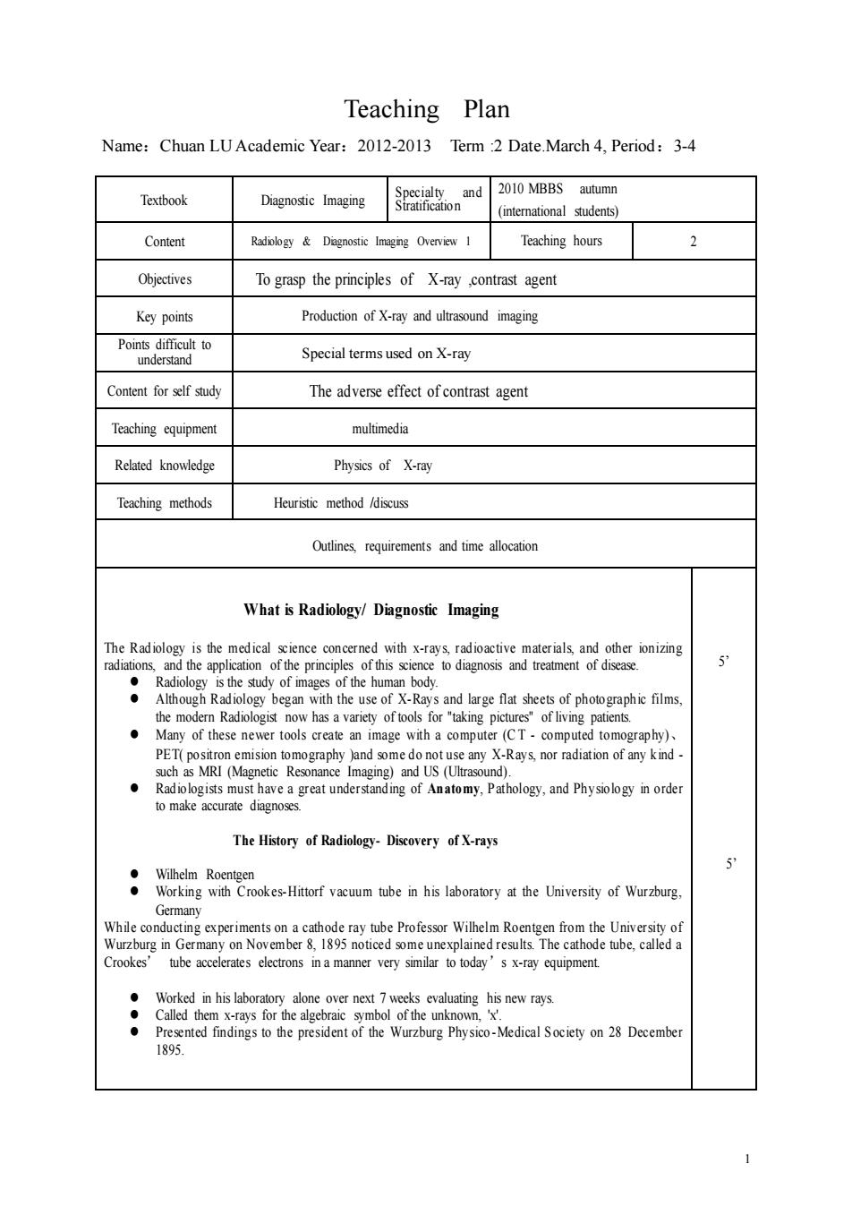
Teaching Plan Name:Chuan LU Academic Year:2012-2013 Term :2 Date.March 4,Period:3-4 2010 MBBS autumn Textbook Diagnosic Imaging onnd mtemationals$tudents Content Teaching hours 2 Objectives To grasp the principles of X-ray contrast agent Key points Production of X-ray and urasound imaging o Special terms used on X-ray Content for self study The adverse effect of contrast agent Teaching equipment multimedia Related knowedge Physics of X-ray Teaching methods Heuristic method /discuss Outlines requirements and time allocation What is Radiology/Diagnostic Imaging The Radiology is the medical sene concerned with x-rays,radioactive materials and other ionizing the modern Radio ave a great understanding of Antmy,Pathology,and Physologyinorder The History of Radiology-Discovery of X-rays :life Loyee时o时u 5 Germany Crookes'tube accelerates electrons in a manner very similar to today's x-ray equipment m时atgsocesdatoftrWbagwsoAksalSocym28Dernic 1 1 Teaching Plan Name:Chuan LU Academic Year:2012-2013 Term :2 Date.March 4, Period:3-4 Textbook Diagnostic Imaging Specialty and Stratification 2010 MBBS autumn (international students) Content Radiology & Diagnostic Imaging Overview 1 Teaching hours 2 Objectives To grasp the principles of X-ray ,contrast agent Key points Production of X-ray and ultrasound imaging Points difficult to understand Special terms used on X-ray Content for self study The adverse effect of contrast agent Teaching equipment multimedia Related knowledge Physics of X-ray Teaching methods Heuristic method /discuss Outlines, requirements and time allocation What is Radiology/ Diagnostic Imaging The Radiology is the medical science concerned with x-rays, radioactive materials, and other ionizing radiations, and the application of the principles of this science to diagnosis and treatment of disease. ⚫ Radiology is the study of images of the human body. ⚫ Although Radiology began with the use of X-Rays and large flat sheets of photographic films, the modern Radiologist now has a variety of tools for "taking pictures" of living patients. ⚫ Many of these newer tools create an image with a computer (C T - computed tomography)、 PET( positron emision tomography )and some do not use any X-Rays, nor radiation of any kind - such as MRI (Magnetic Resonance Imaging) and US (Ultrasound). ⚫ Radiologists must have a great understanding of An atomy, P athology, and Ph ysiology in order to make accurate diagnoses. The History of Radiology- Discovery of X-rays ⚫ Wilhelm Roentgen ⚫ Working with Crookes-Hittorf vacuum tube in his laboratory at the University of Wurzburg, Germany While conducting experiments on a cathode ray tube Professor Wilhelm Roentgen from the University of Wurzburg in Germany on November 8, 1895 noticed some unexplained results. The cathode tube, called a Crookes’ tube accelerates electrons in a manner very similar to today’s x-ray equipment. ⚫ Worked in his laboratory alone over next 7 weeks evaluating his new rays. ⚫ Called them x-rays for the algebraic symbol of the unknown, 'x'. ⚫ Presented findings to the president of the Wurzburg Physico -Medical S ociety on 28 December 1895. 5’ 5’