Teaching Plan Name:Chuan LU Academic Year:2012-2013 Term :2 Date.March 4,Period:3-4 2010 MBBS autumn Textbook Diagnosic Imaging onnd mtemationals$tudents Content Teaching hours 2 Objectives To grasp the principles of X-ray contrast agent Key points Production of X-ray and urasound imaging o Special terms used on X-ray Content for self study The adverse effect of contrast agent Teaching equipment multimedia Related knowedge Physics of X-ray Teaching methods Heuristic method /discuss Outlines requirements and time allocation What is Radiology/Diagnostic Imaging The Radiology is the medical sene concerned with x-rays,radioactive materials and other ionizing the modern Radio ave a great understanding of Antmy,Pathology,and Physologyinorder The History of Radiology-Discovery of X-rays :life Loyee时o时u 5 Germany Crookes'tube accelerates electrons in a manner very similar to today's x-ray equipment m时atgsocesdatoftrWbagwsoAksalSocym28Dernic 1
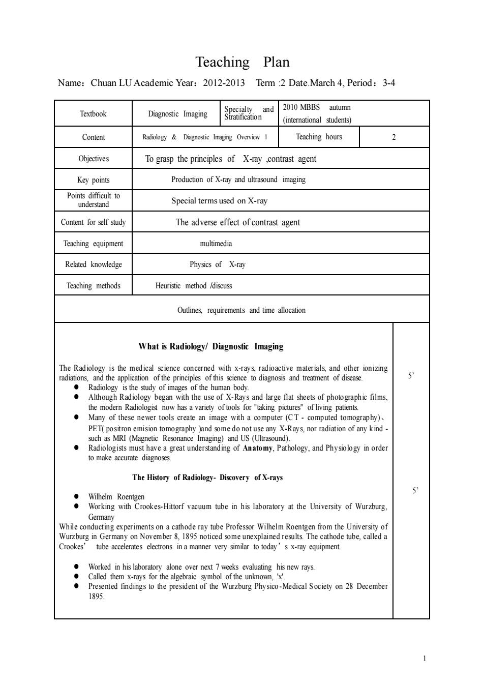
1 Teaching Plan Name:Chuan LU Academic Year:2012-2013 Term :2 Date.March 4, Period:3-4 Textbook Diagnostic Imaging Specialty and Stratification 2010 MBBS autumn (international students) Content Radiology & Diagnostic Imaging Overview 1 Teaching hours 2 Objectives To grasp the principles of X-ray ,contrast agent Key points Production of X-ray and ultrasound imaging Points difficult to understand Special terms used on X-ray Content for self study The adverse effect of contrast agent Teaching equipment multimedia Related knowledge Physics of X-ray Teaching methods Heuristic method /discuss Outlines, requirements and time allocation What is Radiology/ Diagnostic Imaging The Radiology is the medical science concerned with x-rays, radioactive materials, and other ionizing radiations, and the application of the principles of this science to diagnosis and treatment of disease. ⚫ Radiology is the study of images of the human body. ⚫ Although Radiology began with the use of X-Rays and large flat sheets of photographic films, the modern Radiologist now has a variety of tools for "taking pictures" of living patients. ⚫ Many of these newer tools create an image with a computer (C T - computed tomography)、 PET( positron emision tomography )and some do not use any X-Rays, nor radiation of any kind - such as MRI (Magnetic Resonance Imaging) and US (Ultrasound). ⚫ Radiologists must have a great understanding of An atomy, P athology, and Ph ysiology in order to make accurate diagnoses. The History of Radiology- Discovery of X-rays ⚫ Wilhelm Roentgen ⚫ Working with Crookes-Hittorf vacuum tube in his laboratory at the University of Wurzburg, Germany While conducting experiments on a cathode ray tube Professor Wilhelm Roentgen from the University of Wurzburg in Germany on November 8, 1895 noticed some unexplained results. The cathode tube, called a Crookes’ tube accelerates electrons in a manner very similar to today’s x-ray equipment. ⚫ Worked in his laboratory alone over next 7 weeks evaluating his new rays. ⚫ Called them x-rays for the algebraic symbol of the unknown, 'x'. ⚫ Presented findings to the president of the Wurzburg Physico -Medical S ociety on 28 December 1895. 5’ 5’

Outlines requirements and time The Imaging modalities 5 Radiography (XR.Plain Films) raphy (US) (MR) (DSA) ommunication System(PACS) X-Rays 15 Defined:The use of high-energy electromagnetic waves,passing through the body onto a photographic film,to producea picture of the internal structures of the body for diagnosis and herapy Properties ofX-rays 20 aphic effect ogical effec X-Rays High-energy electromagnetic waves an visible light Able topenetrate varying densities Much the xposing aph Imaging Principle of X-rays X-rays pass through tissue to expose film ●Passage depends: s/gram Energy of the Radiation Imaging Principle of X-ray When X-rays traverse through the body,different tissues absorb different amounts of The differential absorption of tissues is the cardinal feature of x-ray image formation
2 Outlines, requirements and time allocation . The Imaging modalities ⚫ Radiography (XR, Plain Films) ⚫ Ultrasonography (US) ⚫ Computed Tomography (CT) ⚫ Magnetic Resonance Imaging (MR) ⚫ Digital Subtraction of Angiography (DSA) ⚫ Nuclear Medicine (PET) ⚫ Picture Archiving and Communication System (PACS ) X-Rays Defined:The use of high-energy electromagnetic waves, passing through the body onto a photographic film, to produce a picture of the internal structures of the body for diagnosis and therapy Properties of X-rays ⚫ Penetration ⚫ Fluorescent effect ⚫ Photographic effect ⚫ Ionizing effect ⚫ Biological effect X-Rays ⚫ High-energy electromagnetic waves ⚫ Travel in straight lines ⚫ Shorter wave length than visible light ⚫ Able to penetrate solid materials of varying densities ⚫ Capable of exposing a photographic plate (x-ray film) Much the same way as a camera exposes film Imaging Principle of X-rays ⚫ X-rays pass through tissue to expose film ⚫ Passage depends: Tissue Character Density Atomic number Number of electrons/gram Thickness (scatter) Energy of the Radiation Imaging Principle of X-ray ⚫ When X-rays traverse through the body, different tissues absorb different amounts of X-rays. ⚫ The unabsorbed X-rays get through to expose the X-ray film and create the image (photographic effect). The differential absorption of tissues is the cardinal feature of x-ray image formation 5’ 15’ 20

Outlines requirements and time allocation 15 Fat and Lipid : 。Heavy Metal Synonym:High density InChinese高密度 度 Radiopaque Synonym:High density 20° Soft tissue and body fluid(water density) ht d posion Radiolucent ic cae mediastinum,abdominal area(light or white) Synonym:High density Methods of X-ray Examination ·Conventional: roscopy : aphy X-RAY:Normal abdomen APview ofabdomen in supine position Low dem bowel gas ·Bones.:High density All abdominal 10 · compounds into the vessels.etc. agents 3
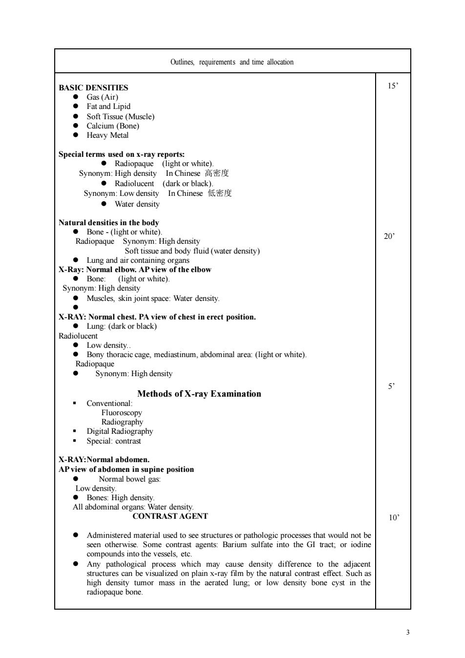
3 Outlines, requirements and time allocation BASIC DENSITIES ⚫ Gas (Air) ⚫ Fat and Lipid ⚫ Soft Tissue (Muscle) ⚫ Calcium (Bone) ⚫ Heavy Metal Special terms used on x-ray reports: ⚫ Radiopaque (light or white). Synonym: High density In Chinese 高密度 ⚫ Radiolucent (dark or black). Synonym: Low density In Chinese 低密度 ⚫ Water density Natural densities in the body ⚫ Bone - (light or white). Radiopaque Synonym: High density Soft tissue and body fluid (water density) ⚫ Lung and air containing organs X-Ray: Normal elbow. AP view of the elbow ⚫ Bone: (light or white). Synonym: High density ⚫ Muscles, skin joint space: Water density. ⚫ X-RAY: Normal chest. PA view of chest in erect position. ⚫ Lung: (dark or black) Radiolucent ⚫ Low density. ⚫ Bony thoracic cage, mediastinum, abdominal area: (light or white). Radiopaque ⚫ Synonym: High density Methods of X-ray Examination ▪ Conventional: Fluoroscopy Radiography ▪ Digital Radiography ▪ Special: contrast X-RAY:Normal abdomen. AP view of abdomen in supine position ⚫ Normal bowel gas: Low density. ⚫ Bones: High density. All abdominal organs: Water density. CONTRAST AGENT ⚫ Administered material used to see structures or pathologic processes that would not be seen otherwise. Some contrast agents: Barium sulfate into the GI tract; or iodine compounds into the vessels, etc. ⚫ Any pathological process which may cause density difference to the adjacent structures can be visualized on plain x-ray film by the natural contrast effect. Such as high density tumor mass in the aerated lung; or low density bone cyst in the radiopaque bone. 15’ 20’ 5’ 10’

Outlinesrequrements and time The pathological tissue which has almost the same density as the adjacent structures cannot be seen on plain film. Artificial contrast is needed to create a density difference CONTRAST RADIOGRAPHY(Never say“Dye”) Barium studies(BAS,UGI,LGI/BE) ● (Percutaneous Transhepatic/ravenous) Urography(Ki ney function)(IV) ● 的een rography X-RAY: AD rmal IVP and ftered and excreted by the kidneys. injected intravenously Barium enema AP view of a m outlining the lumen of the large bowel (high density-) X-Rav Upper gastrointestinal tract study Normal AP supine view of the abdomen following the ora administration of barium
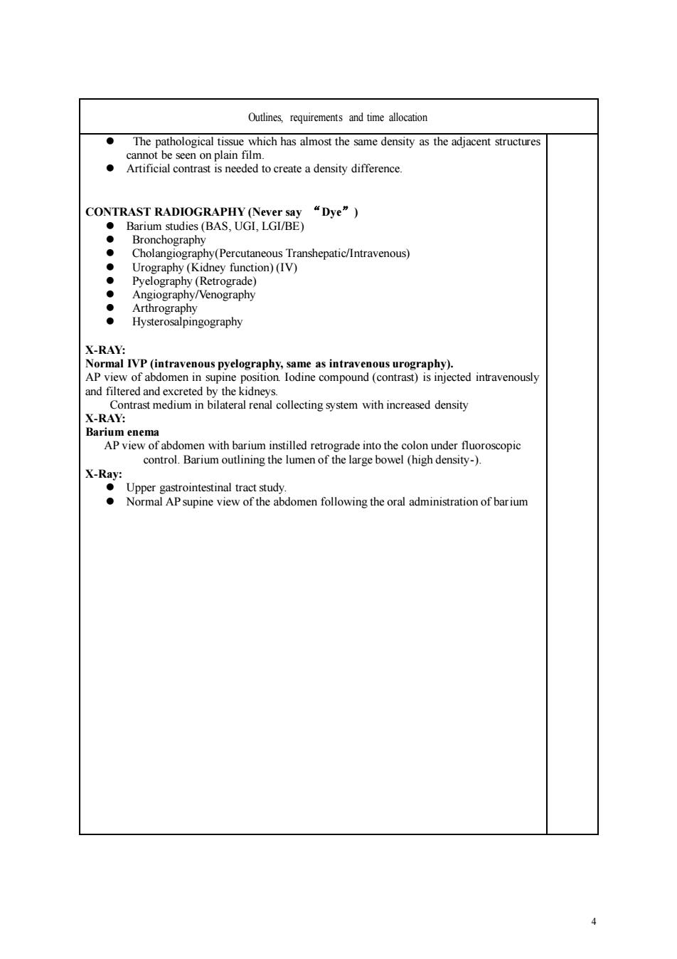
4 Outlines, requirements and time allocation ⚫ The pathological tissue which has almost the same density as the adjacent structures cannot be seen on plain film. ⚫ Artificial contrast is needed to create a density difference. CONTRAST RADIOGRAPHY (Never say “Dye”) ⚫ Barium studies (BAS, UGI, LGI/BE) ⚫ Bronchography ⚫ Cholangiography(Percutaneous Transhepatic/Intravenous) ⚫ Urography (Kidney function) (IV) ⚫ Pyelography (Retrograde) ⚫ Angiography/Venography ⚫ Arthrography ⚫ Hysterosalpingography X-RAY: Normal IVP (intravenous pyelography, same as intravenous urography). AP view of abdomen in supine position. Iodine compound (contrast) is injected intravenously and filtered and excreted by the kidneys. Contrast medium in bilateral renal collecting system with increased density X-RAY: Barium enema AP view of abdomen with barium instilled retrograde into the colon under fluoroscopic control. Barium outlining the lumen of the large bowel (high density-). X-Ray: ⚫ Upper gastrointestinal tract study. ⚫ Normal AP supine view of the abdomen following the oral administration of barium