Teaching Plan Name:Linxiang Liu Academic Year:2012-2013 Term:2 Date:May7 Period:3-4 2010 MBBS autumn Textbook Diagnostic Imaging Suaion (students) Content Pulmonary inflammatory disease Teaching 2 Objectives Pulmonary inflammatory disease Key points Lobar pneumonia,Bronchopneumonia,Lung abscess Points difficult to Lobar pneumonia,Lung abscess Content for self study CT、MR anatomy Teaching equipment multimedia Related Medical imaging technique,anatomy,pathology,medicine,surgery knowledge Teaching methods Heuristic method discuss Outlines,requirements and time allocation atory Disease r pneumonia ● young adults,pathologically divided in four stages but radiologically 20 divided 3 stages,because x-ray exam can not differentiate red-liver stage from gray eoaimbRaenspon8e consolidation loss its homogeneity,the residual strands and strips may be mistaken for fibrosis if patients history are not considered. Bronchopneumonia 15 Commonly seen in infants or teenagers.In pathology it involves acute inflammation of multiple bronchioles,character by exudation and bronchiole obstruction by secretion. in lob Lung abscess 20 primary hematogeou :patch or mass like shadow.After discharge of necrosis,i cavity.usually with thick wall,irregular linings,with or without air-fluid 0
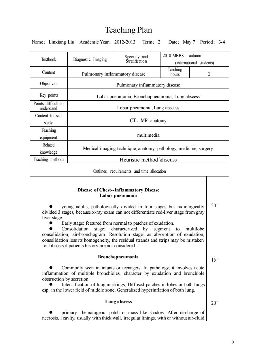
0 Teaching Plan Name:Linxiang Liu Academic Year:2012-2013 Term:2 Date:May 7 Period:3-4 Textbook Diagnostic Imaging Specialty and Stratification 2010 MBBS autumn (international students) Content Pulmonary inflammatory disease Teaching hours 2 Objectives Pulmonary inflammatory disease Key points Lobar pneumonia, Bronchopneumonia, Lung abscess Points difficult to understand Lobar pneumonia, Lung abscess Content for self study CT、MR anatomy Teaching equipment multimedia Related knowledge Medical imaging technique, anatomy, pathology, medicine, surgery Teaching methods Heuristic method \discuss Outlines, requirements and time allocation Disease of Chest-Inflammatory Disease Lobar pneumonia ⚫ young adults, pathologically divided in four stages but radiologically divided 3 stages, because x-ray exam can not differentiate red-liver stage from gray liver stage. ⚫ Early stage: featured from normal to patches of exudation. ⚫ Consolidation stage: characterized by segment to multilobe consolidation, air-bronchogram. Resolution stage: as absorption of exudation, consolidation loss its homogeneity, the residual strands and strips may be mistaken for fibrosis if patients history are not considered. Bronchopneumonia ⚫ Commonly seen in infants or teenagers. In pathology, it involves acute inflammation of multiple bronchioles, character by exudation and bronchiole obstruction by secretion. ⚫ Intensification of lung markings, Diffused patches in lobes or both lungs esp. in the lower field of middle zone, Generalized hyperinflation of both lung. Lung abscess ⚫ primary hematogeou :patch or mass like shadow. After discharge of necrosis, i cavity, usually with thick wall, irregular linings, with or without air-fluid 20’ 15’ 20’

which Hematogeous abscess usually is the result of septicemia,it appears as multiple tube-like dilati inal bronchi Plainm may varesfo norma toocdesification of lung markings.There may be exudation or air-fluid level if the foci are involve ronchography used to be the only way tha exten lung and raphy in the diagnosis of bro Lung Tuberculosis TYP Primary TB.Hematogenous TB,Infiltrative TB,Chronic fibro-cavitative TB,Tuberculosis pleuritis Primary TB 尔 ● ening Draining lymph nodes may enlarge as part of this sensitivity reaction ● Radiography of the chest in primary tuberculosis varies from a small subpleural ● Enlarged lymph nodes cause distal atelectasis Pleural effusion is not an unusual feature Lung changes take months or years to clear,and any calcified lesions persist indefinitely Hematogenous TB Chronic dissemination of TB 10 Miliary tuberculosis: ● Chest film demonstrates multiple,small nodular opacities throughout both lungs that are too numerous to count Infiltrative TB 15 upper lung predilection,Tuberculoma:sharp margin,solitary/multiple. regular/irregular,calcification,"Satellite"foci.Cavitation:Vary in walls and with the acitveness of disease.Patchies and monia ●
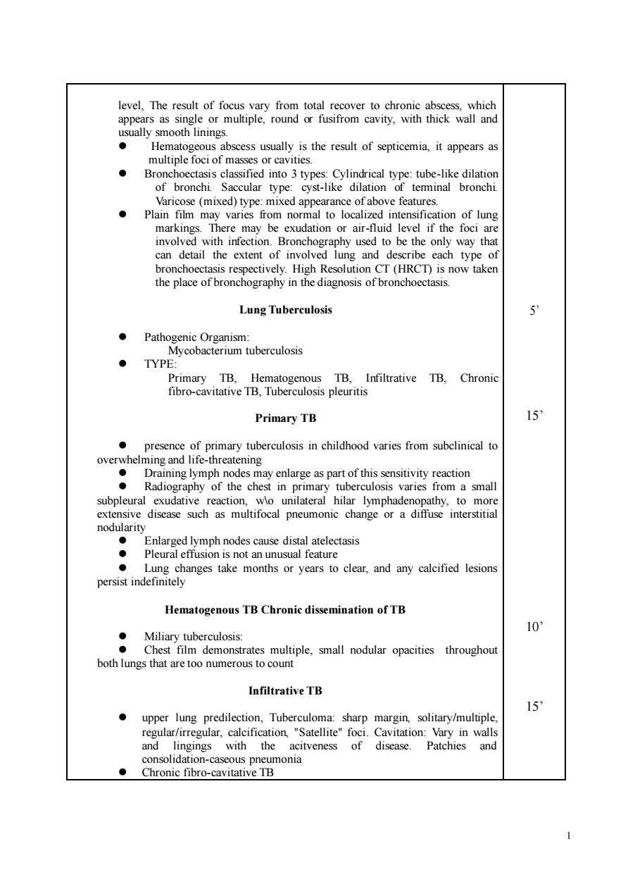
1 level, The result of focus vary from total recover to chronic abscess, which appears as single or multiple, round or fusifrom cavity, with thick wall and usually smooth linings. ⚫ Hematogeous abscess usually is the result of septicemia, it appears as multiple foci of masses or cavities. ⚫ Bronchoectasis classified into 3 types: Cylindrical type: tube-like dilation of bronchi. Saccular type: cyst-like dilation of terminal bronchi. Varicose (mixed) type: mixed appearance of above features. ⚫ Plain film may varies from normal to localized intensification of lung markings. There may be exudation or air-fluid level if the foci are involved with infection. Bronchography used to be the only way that can detail the extent of involved lung and describe each type of bronchoectasis respectively. High Resolution CT (HRCT) is now taken the place of bronchography in the diagnosis of bronchoectasis. Lung Tuberculosis ⚫ Pathogenic Organism: Mycobacterium tuberculosis ⚫ TYPE: Primary TB, Hematogenous TB, Infiltrative TB, Chronic fibro-cavitative TB, Tuberculosis pleuritis Primary TB ⚫ presence of primary tuberculosis in childhood varies from subclinical to overwhelming and life-threatening ⚫ Draining lymph nodes may enlarge as part of this sensitivity reaction ⚫ Radiography of the chest in primary tuberculosis varies from a small subpleural exudative reaction, w\o unilateral hilar lymphadenopathy, to more extensive disease such as multifocal pneumonic change or a diffuse interstitial nodularity ⚫ Enlarged lymph nodes cause distal atelectasis ⚫ Pleural effusion is not an unusual feature ⚫ Lung changes take months or years to clear, and any calcified lesions persist indefinitely Hematogenous TB Chronic dissemination of TB ⚫ Miliary tuberculosis: ⚫ Chest film demonstrates multiple, small nodular opacities throughout both lungs that are too numerous to count Infiltrative TB ⚫ upper lung predilection, Tuberculoma: sharp margin, solitary/multiple, regular/irregular, calcification, "Satellite" foci. Cavitation: Vary in walls and lingings with the acitveness of disease. Patchies and consolidation-caseous pneumonia ⚫ Chronic fibro-cavitative TB 5’ 15’ 10’ 15’