
TAISHANMEDICAL UNIVERSITY School of Radiology SETA 6Semester Final Exams,September Session 2009 Time)-30 minutes Full Marks-80 Name. Roll No. Tick mark (v)the one best answer 1.Pneumomediatinum refersto the presence of air in a.Neck b.Around the great vessels C. Pleural cavity d.Abdominal cavity 2.Retropharyngeal abscess is characterised by 6 Involves the prevertebral apace 3.Punched t lytic lesior b.Hyperparathyroidism c.Metastases d.None of the above 4.The cranial nervereadily visualised on CT images a.1 b.II ty of choice ntra 8osngaeutesutbartachnoidhemorge ntra st en IMR d Non co ntrast MR 6.All are true about intracranial hematomasexcept a.Acute hematoma appear hyperdense on CT b.Extradural hematomaappears as a lenticular shaped extra-axial collection c.Acute subdural hemorraghe appears as hyperdense in sulcal spaces&basal cisterns d.The commonest site for hypertensive bleed is basal ganglia 7.Which of the following foreign bodies can be visualized radiographically 8.Absenc ga s on abdominal radiograph suggests 。D al SBO b.Pso as ahs c.Chest infection d.Midgut volvulus 9.Contrast media of choice in investigating a suspected case of ileal perforation a.Barium sulphate b.Gastrograffin
Taishan medical university School of Radiology SET A 6 th Semester Final Exams, September Session 2009 Time)– 30 minutes Full Marks-30 Name. Roll No. . Tick mark (√) the one best answer 1. Pneumomediatinum refers to the presence of air in a. Neck b. Around the great vessels c. Pleural cavity d. Abdominal cavity 2. Retropharyngeal abscess is characterised by a. Involves the prevertebral apace b. Best seen on lateral x-ray c. May occur with TB of cervical verterbrae d. All the above 3. Punched out lytic lesions in the skull are characteristic of a. Multiple myeloma b. Hyperparathyroidism c. Metastases d. None of the above 4. The cranial nerve readily visualised on CT images a. I b. II c. III d. IV 5. Modality of choice diagnosing acute subarachnoid hemorraghe a. Non contrast CT b. Contrast enhanced CT c. Contrast enhanced MR d. Non contrast MR 6. All are true about intracranial hematomas except a. Acute hematoma appear hyperdense on CT b. Extradural hematoma appears as a lenticular shaped extra-axial collection c. Acute subdural hemorraghe appears as hyperdense in sulcal spaces & basal cisterns d. The commonest site for hypertensive bleed is basal ganglia 7. Which of the following foreign bodies can be visualized radiographically a. Glass b. Wood c. Plastic d. None 8. Absence of gas on abdominal radiograph suggests a. Proximal SBO b. Psoas abscess c. Chest infection d. Midgut volvulus 9. Contrast media of choice in investigating a suspected case of ileal perforation a. Barium sulphate b. Gastrograffin
c.Dionosul viscous d Linoidol 10.Intravenous urography is contraindicated in all except a.Pregnancy b. Renal insufficiency :d Renal trauma Hypersensitivity to contrast 11.Best investigation in acute cholecystitisis a. echnetium scan lain x ray abd 12 Left atrial hy radiologically as Double cardiac h Left bronchial elevation Straightnening of left border d.all of the above 13.Left to right shunt is usually demonstrated in chest xray by a.Increase in pulmonary venous markings b. Increase in size of pulmonary arteries Increase in pulmonary vascular markings 14.Notchingof ribs are seenin p.ASD teins anmoly d.Coarctation of aorta 15.lleum in barium looks like a Characteristic b.Irregular dilatation c.Characterless d.None of the above 16.Oligaemiclung fields are seen in a.T0时 17.Angle No e of the trach hrsiamisincreasedinenlargemento c.Rt atrium d It atrium 18.Free air under the diaphragm may be seen in all except a.Peritoneal dialysis b. Bowel perforation Immediate post operative 19.nvestigation of chodiagnosingrenal calculiis d 20.Bats wingappearanceon chest skiagram is classically seenin cases of y pulmonary HTN b Pulmor ary oedema c.Pericardial effusion
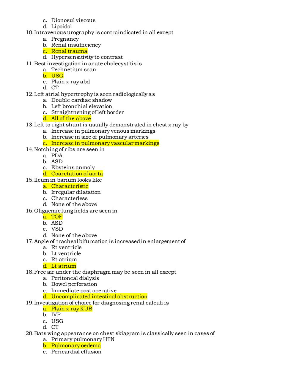
c. Dionosul viscous d. Lipoidol 10.Intravenous urography is contraindicated in all except a. Pregnancy b. Renal insufficiency c. Renal trauma d. Hypersensitivity to contrast 11.Best investigation in acute cholecystitis is a. Technetium scan b. USG c. Plain x ray abd d. CT 12.Left atrial hypertrophy is seen radiologically as a. Double cardiac shadow b. Left bronchial elevation c. Straightnening of left border d. All of the above 13.Left to right shunt is usually demonstrated in chest x ray by a. Increase in pulmonary venous markings b. Increase in size of pulmonary arteries c. Increase in pulmonary vascular markings 14.Notching of ribs are seen in a. PDA b. ASD c. Ebsteins anmoly d. Coarctation of aorta 15.Ileum in barium looks like a. Characteristic b. Irregular dilatation c. Characterless d. None of the above 16.Oligaemic lung fields are seen in a. TOF b. ASD c. VSD d. None of the above 17.Angle of tracheal bifurcation is increased in enlargement of a. Rt ventricle b. Lt ventricle c. Rt atrium d. Lt atrium 18.Free air under the diaphragm may be seen in all except a. Peritoneal dialysis b. Bowel perforation c. Immediate post operative d. Uncomplicated intestinal obstruction 19.Investigation of choice for diagnosing renal calculi is a. Plain x ray KUB b. IVP c. USG d. CT 20.Bats wing appearance on chest skiagram is classically seen in cases of a. Primary pulmonary HTN b. Pulmonary oedema c. Pericardial effusion
d.All of the above 21.Ga in medici a.Measuring the radioactivity is used for c.Monitoring the surface contamination b.Organ imaging d.Radioimmuneassay 22.The cranial nerve readily visualized on CT images 02 Al about intr xcep c.Acute subdural hemorraghe appears as b.Extradural hemato na appears as a hyperdense in sulcal spaces basal cisterns lenticular shaped extra-axial collection d.The com onest site for hypertensive bleed is I ganglia graphically h Wood None 25.Contrast media of choice in investigating a suspected case of ileal perforation a.Barium sulphate C. Dionosul viscous 26.Angl astrogr Lipoidol ifurcation is increased in enlargement of b Lt ventricle d.Lt atrium 27.Following renal calculi are radiopaque except a.Uric acid Triple phosphate Calciun xalat d.Staghorn calculus 28.Rad otection shields are made up of d.Tead c. 29.Which radiations is most harmful to body Beta particl d.am na rays who a.Diagnoses the disease using the imaging moda e like e a physician
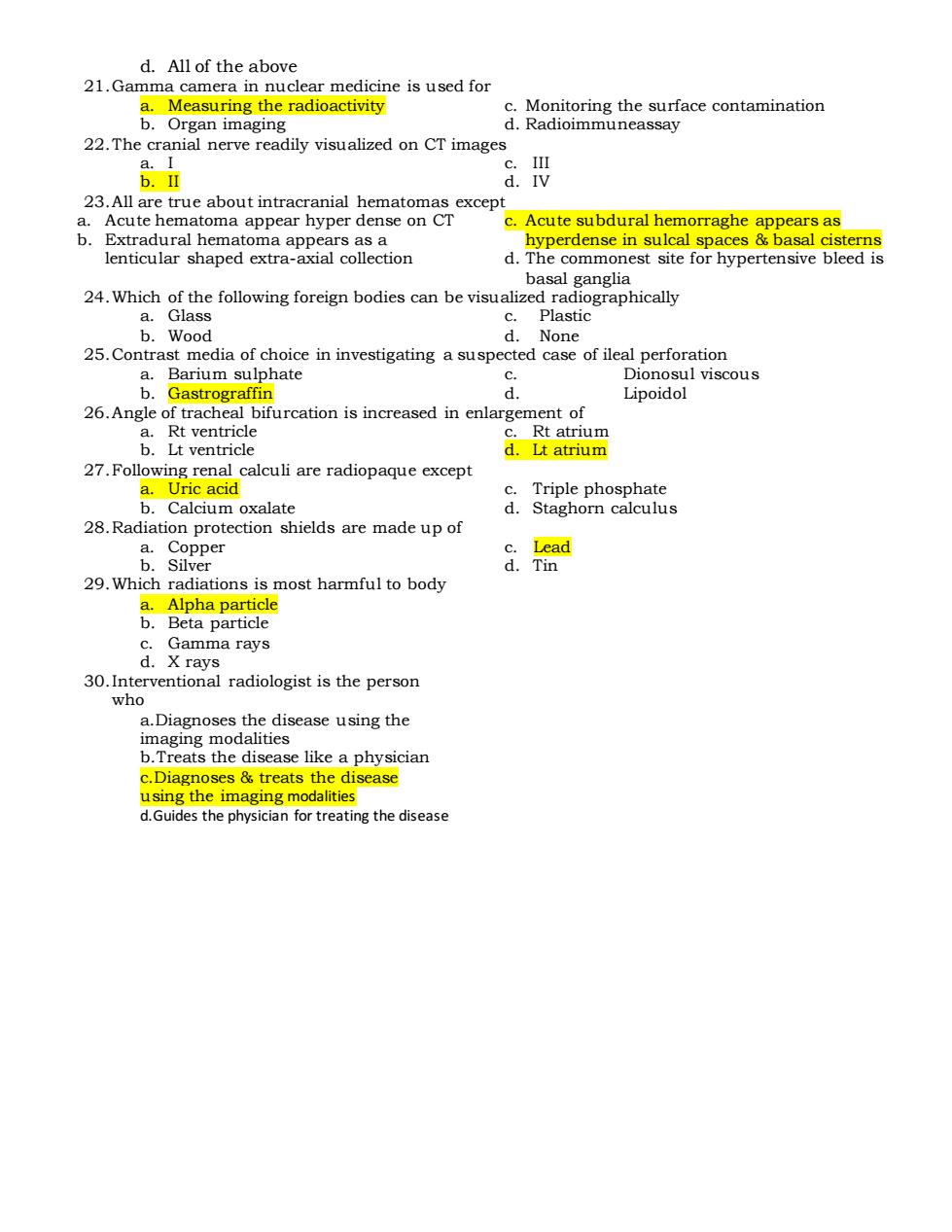
d. All of the above 21.Gamma camera in nuclear medicine is used for a. Measuring the radioactivity b. Organ imaging c. Monitoring the surface contamination d. Radioimmuneassay 22.The cranial nerve readily visualized on CT images a. I b. II c. III d. IV 23.All are true about intracranial hematomas except a. Acute hematoma appear hyper dense on CT b. Extradural hematoma appears as a lenticular shaped extra-axial collection c. Acute subdural hemorraghe appears as hyperdense in sulcal spaces & basal cisterns d. The commonest site for hypertensive bleed is basal ganglia 24.Which of the following foreign bodies can be visualized radiographically a. Glass b. Wood c. Plastic d. None 25.Contrast media of choice in investigating a suspected case of ileal perforation a. Barium sulphate b. Gastrograffin c. Dionosul viscous d. Lipoidol 26.Angle of tracheal bifurcation is increased in enlargement of a. Rt ventricle b. Lt ventricle c. Rt atrium d. Lt atrium 27.Following renal calculi are radiopaque except a. Uric acid b. Calcium oxalate c. Triple phosphate d. Staghorn calculus 28.Radiation protection shields are made up of a. Copper b. Silver c. Lead d. Tin 29.Which radiations is most harmful to body a. Alpha particle b. Beta particle c. Gamma rays d. X rays 30.Interventional radiologist is the person who a.Diagnoses the disease using the imaging modalities b.Treats the disease like a physician c.Diagnoses & treats the disease using the imaging modalities d.Guides the physician for treating the disease
TAISHAN MEDICAL UNIVERSITY Schodl of Radiology SETA 6"Semester Final Exams,September Session 2009 Time -2.30 houn Full Marks70 Date-12 January,2010 Pass marks -42 Attempt all questions 1.What are the types of ultrasound transducer?How do you choose a propertransducer when linear array transducer,sector transducer.curved array transducer and special transducers The type of transducer selected for a particular examination depends upon several factors The type of examination: The echocardiographic examination is performed with a sector transducer(3.5MHz).Obstetric and gynecologic scans are usually performed with a curved or linear array transducer(3.5MHz). The size of the patient ●For children:1oMHz Special transducers have been produced to help view specific areas 2 Describe CT features of Cerebral Infarction Three phases may be identified:acute,subacute and chronic In the acute phase:about 60%of scans may be considered normal within the first fewhours some subtle early signs may be detected loss of grey-white matter interface >obscuration of the lentiform nucleus hyperdensity of the middle cerebral artery A rapid development of the picture may be seen after 6-8 hours with the development of cytotoxic oedema.the infarcted area becomes markedly hypodense with involvement of both grey and white matter,usually in a wedge-shaped area comresponding to the region of distribution of the occluded vessel about 15-20%of cases haemorrhagic foci may be detected after 24-48 hours 3.Describe the imaging features of Meningioma. .CT:shows a well marginated oval or lobulated mass.attached to the dura at an obtuse angle.Most lesions are homogeneously hyperdense,some isodense and a few hypodense ReuTiZie Pou
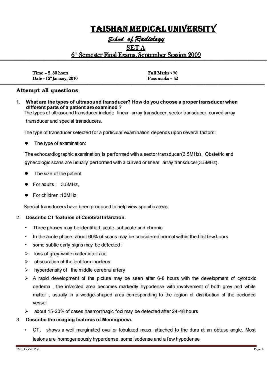
Ren Ti Zie Pou. Page 4 Taishan medical university School of Radiology SET A 6 th Semester Final Exams, September Session 2009 Time – 2. 30 hours Full Marks- 70 Date– 12th January, 2010 Pass marks – 42 Attempt all questions 1. What are the types of ultrasound transducer? How do you choose a proper transducer when different parts of a patient are examined ? The types of ultrasound transducer include linear array transducer, sector transducer ,curved array transducer and special transducers. The type of transducer selected for a particular examination depends upon several factors: ⚫ The type of examination: The echocardiographic examination is performed with a sector transducer(3.5MHz). Obstetric and gynecologic scans are usually performed with a curved or linear array transducer(3.5MHz). ⚫ The size of the patient ⚫ For adults : 3.5MHz, ⚫ For children :10MHz Special transducers have been produced to help view specific areas. 2. Describe CT features of Cerebral Infarction. • Three phases may be identified: acute, subacute and chronic • In the acute phase :about 60% of scans may be considered normal within the first few hours • some subtle early signs may be detected : ➢ loss of grey-white matter interface ➢ obscuration of the lentiform nucleus ➢ hyperdensity of the middle cerebral artery ➢ A rapid development of the picture may be seen after 6-8 hours with the development of cytotoxic oedema , the infarcted area becomes markedly hypodense with involvement of both grey and white matter , usually in a wedge-shaped area corresponding to the region of distribution of the occluded vessel ➢ about 15-20% of cases haemorrhagic foci may be detected after 24-48 hours 3. Describe the imaging features of Meningioma. • CT: shows a well marginated oval or lobulated mass, attached to the dura at an obtuse angle. Most lesions are homogeneously hyperdense, some isodense and a few hypodense
Calcifications are diffuse or focal Marked homogeneous enhancement MRI:Isointensity on both T1WI and T2WI Sixty percent of meningiomas have a"dural tail of enhancement Marked homogeneous enhancement 4.(a)What are the differences between radiological findings in pulmonary malignant nodules&Benign nodules? Pulmonary malignant nodules often appear ill-defined and/or irregular in shape. Benign nodules are often well-defined,round and smooth in contour. Air bronchograms within a nodule are more common in malignancies than in benign nodules Cavitation is also more common in malignant nodules than benign nodules (b)Barium examination includes four kinds of imagings,justwrite down their names Mucosa imaging Turgor(full)imaging Compression imaging Double contrast imaging 5.Describe the ultrasound and CT findings of hepatocellular carcinoma. Ultrasound findings: HyperechoicHCC(13%)due to fatty metamophosis or marked dilatation of sinusoids Hypoechoic HCC(26%)due to solid tumor Mixed echogenicity (6%due to nonliquefactive tumor necrosis Portal vein invasion (25-48%) CT findings: Unenhanced CT: hypodense mass/rarely isodense/hyperdense in fatty live Biphasic Contrast Enhanced CT: enhancement during hepatic arterial phase(80%) decreased attenuation during portal venous phase with inhomogeneous areas of contrast accumulation isodensity on delayed scans(10%) thin contrast-enhancing capsule(50%)due to rapid washout wedge-shaped areas of decreased attenuation(segmental/lobar perfusion defects due portal vein occlusion by tumor thrombus) 6.What are the advantages&disadvantage of various modalities in biliary imaging? Modality Advantages Disadvantages ERCP Permits simultaneous biopsy or Invasive.may not be possible due to altered anatomy Treatment following surgery RenTiZe Pou
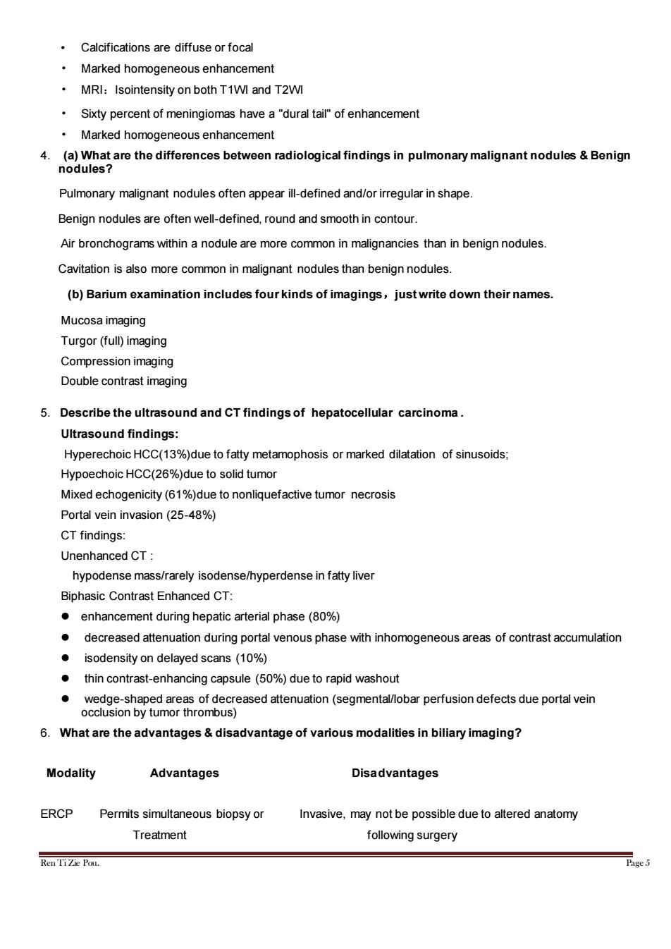
Ren Ti Zie Pou. Page 5 • Calcifications are diffuse or focal • Marked homogeneous enhancement • MRI:Isointensity on both T1WI and T2WI • Sixty percent of meningiomas have a "dural tail" of enhancement • Marked homogeneous enhancement 4. (a) What are the differences between radiological findings in pulmonary malignant nodules & Benign nodules? Pulmonary malignant nodules often appear ill-defined and/or irregular in shape. Benign nodules are often well-defined, round and smooth in contour. Air bronchograms within a nodule are more common in malignancies than in benign nodules. Cavitation is also more common in malignant nodules than benign nodules. (b) Barium examination includes four kinds of imagings,just write down their names. Mucosa imaging Turgor (full) imaging Compression imaging Double contrast imaging 5. Describe the ultrasound and CT findings of hepatocellular carcinoma . Ultrasound findings: Hyperechoic HCC(13%)due to fatty metamophosis or marked dilatation of sinusoids; Hypoechoic HCC(26%)due to solid tumor Mixed echogenicity (61%)due to nonliquefactive tumor necrosis Portal vein invasion (25-48%) CT findings: Unenhanced CT : hypodense mass/rarely isodense/hyperdense in fatty liver Biphasic Contrast Enhanced CT: ⚫ enhancement during hepatic arterial phase (80%) ⚫ decreased attenuation during portal venous phase with inhomogeneous areas of contrast accumulation ⚫ isodensity on delayed scans (10%) ⚫ thin contrast-enhancing capsule (50%) due to rapid washout ⚫ wedge-shaped areas of decreased attenuation (segmental/lobar perfusion defects due portal vein occlusion by tumor thrombus) 6. What are the advantages & disadvantage of various modalities in biliary imaging? Modality Advantages Disadvantages ERCP Permits simultaneous biopsy or Invasive, may not be possible due to altered anatomy Treatment following surgery
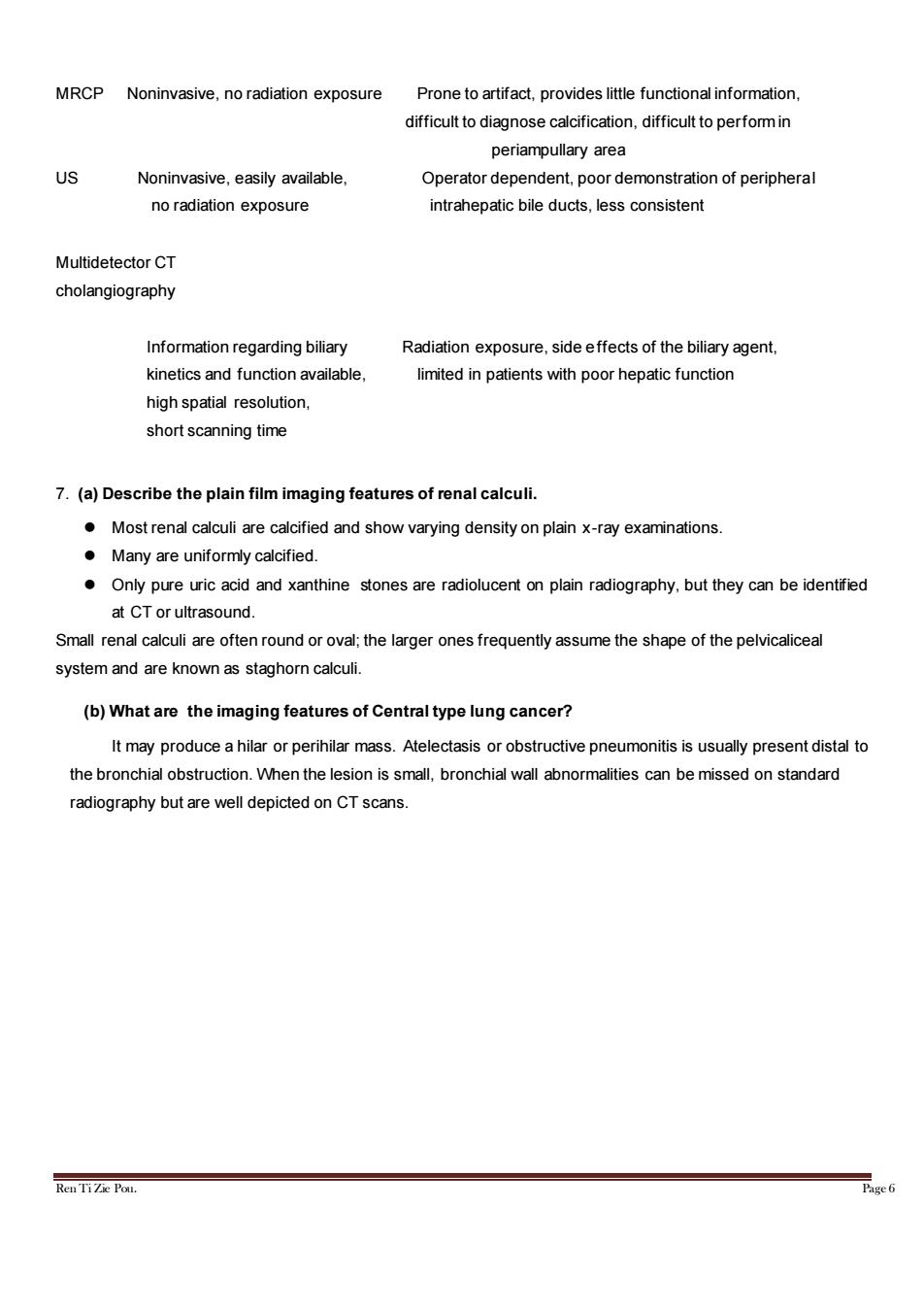
MRCP Noninvasive,no radiation exposure Prone to artifact,provides little functional information, difficult to diagnose calcification,difficult to perform in periampullary area US Noninvasive,easily available, Operator dependent,poor demonstration of peripheral no radiation exposure intrahepatic bile ducts,less consistent Multidetector CT cholangiography Information regarding biliary Radiation exposure,side effects of the biliary agent, kinetics and function available, limited in patients with poor hepatic function high spatial resolution, short scanning time 7.(a)Describe the plain film imaging features of renal calculi. Most renal calculi are calcified and show varying density on plain x-ray examinations. Many are uniformly calcified. Only pure uric acid and xanthine stones are radiolucent on plain radiography,but they can be identified at CT or ultrasound. Small renal calculi are often round or oval;the larger ones frequently assume the shape of the pelvicaliceal system and are known as staghorn calculi. (b)What are the imaging features of Central type lung cancer? It may produce a hilar or perihilar mass.Atelectasis or obstructive pneumonitis is usually present distal to the bronchial obstruction.When the lesion is small,bronchial wall abnormalities can be missed on standard radiography but are well depicted on CT scans. Ren TiZie Pou. Page 6
Ren Ti Zie Pou. Page 6 MRCP Noninvasive, no radiation exposure Prone to artifact, provides little functional information, difficult to diagnose calcification, difficult to perform in periampullary area US Noninvasive, easily available, Operator dependent, poor demonstration of peripheral no radiation exposure intrahepatic bile ducts, less consistent Multidetector CT cholangiography Information regarding biliary Radiation exposure, side effects of the biliary agent, kinetics and function available, limited in patients with poor hepatic function high spatial resolution, short scanning time 7. (a) Describe the plain film imaging features of renal calculi. ⚫ Most renal calculi are calcified and show varying density on plain x-ray examinations. ⚫ Many are uniformly calcified. ⚫ Only pure uric acid and xanthine stones are radiolucent on plain radiography, but they can be identified at CT or ultrasound. Small renal calculi are often round or oval; the larger ones frequently assume the shape of the pelvicaliceal system and are known as staghorn calculi. (b) What are the imaging features of Central type lung cancer? It may produce a hilar or perihilar mass. Atelectasis or obstructive pneumonitis is usually present distal to the bronchial obstruction. When the lesion is small, bronchial wall abnormalities can be missed on standard radiography but are well depicted on CT scans