Teaching Plan Name:Bin FU Academic Year2012-2013 Term:Two Date:June 10 Period:5-6 8e8 2010 MBBS Autumn Textbook Diagnostic Imaging Imaging techniques and normal images for Content 2 Genitourinary system Objectives Introduction of the imaging techniques used in Genitourinary system Key points IVU,CT Image CT and IVU image of kidney Content for self study CT、MRI anatomy Teaching equipment multimedia Related knowledge Medical imaging technique.anatomy,pathology.medicine.surgery Teaching methods Heuristic method \discuss Outlines,requirements and time allocation KUB plain film 10 K-Kidney U-Ureter B-Bladder Identify all calcificationsmajor cause is urinary cakuli Look at the other structures on the film-such as the bone, greater psoas muscle Kidneys:Positions-T12-L3;The left kidney is usually higher than the 。Renal ouis The renal lengths-The normal length of the adult kidney is between 10 and 16cm IVU (intravenous urogram) 10 ultrasound CT/MRI 0
0 Teaching Plan Name:Bin FU Academic Year2012-2013 Term:Two Date:June 10 Period:5~6 Textbook Diagnostic Imaging Specialty and Stratification 2010 MBBS Autumn (international students) Content Imaging techniques and normal images for Genitourinary system Teaching hours 2 Objectives Introduction of the imaging techniques used in Genitourinary system Key points IVU,CT Image Points difficult to understand CT and IVU image of kidney Content for self study CT、MRI anatomy Teaching equipment multimedia Related knowledge Medical imaging technique, anatomy, pathology, medicine, surgery Teaching methods Heuristic method \discuss Outlines, requirements and time allocation KUB plain film ⚫ K-Kidney U-Ureter B-Bladder Identify all calcifications—major cause is urinary calculi Look at the other structures on the film—such as the bone, greater psoas muscle ⚫ Kidneys:Positions—T12~L3;The left kidney is usually higher than the right. ⚫ Renal outlines ⚫ The renal lengths —The normal length of the adult kidney is between 10 and 16cm IVU (intravenous urogram) ⚫ IVP (intravenous pyelography) ⚫ Excretory urography ⚫ The IVU as a standard imaging technique has now been largely replaced by ultrasound/CT/MRI 10’ 10’

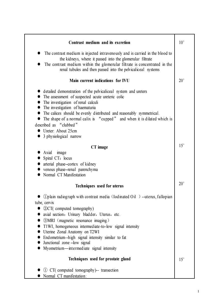
Contrast medium and its excretion 10 The contrast medium is injected intravenously and is carried in the blood to the kidneys,where it passed into the glomerular filtrate The contrast medium within the glomerular filtrate is concentrated in the renal tubules and then passed into the pelvicaliceal systems Main current indications for IVU 20 The investigation ofrenal calculi The investigation of haematuria The calices should be evenly distributed and reasonably symmetrical. ●The shape of a normal calix is“cupped”and when it is dilated which is “clubbed” ●Ureter.About25cm 3 physiological namrow CT image 15 ●Axial image 。Spiral CT:locus aterial phasecortex of kiney venous phase-renal parenchyma Normal CT Manifestation Techniques used for uterus 20 plain radiograph with contrast media (lodinated Oil )-uterus,fallopiar tube.cervix ●②CT(computed tomography) axial section:Urinary bladder,Uterus,etc. ·③MRl(magnetic resonance imaging) TIWI,homogeneous intermediate-to-low signal intensity Uterine Zonal Anatomy on T2WI Endometrium-high signal intensity similar to fat Junctional zone-low signal Myometrium-intermediate signal intensity Techniques used for prostate gland 15 ·①CT(computed tomography-transection Normal CT manifestation:
1 Contrast medium and its excretion ⚫ The contrast medium is injected intravenously and is carried in the blood to the kidneys, where it passed into the glomerular filtrate ⚫ The contrast medium within the glomerular filtrate is concentrated in the renal tubules and then passed into the pelvicaliceal systems Main current indications for IVU ⚫ detailed demonstration of the pelvicaliceal system and ureters ⚫ The assessment of suspected acute ureteric colic ⚫ The investigation of renal calculi ⚫ The investigation of haematuria ⚫ The calices should be evenly distributed and reasonably symmetrical. ⚫ The shape of a normal calix is “cupped” and when it is dilated which is described as “clubbed” ⚫ Ureter: About 25cm ⚫ 3 physiological narrow CT image ⚫ Axial image ⚫ Spiral CT:locus ⚫ arterial phase-cortex of kidney ⚫ venous phase-renal parenchyma ⚫ Normal CT Manifestation Techniques used for uterus ⚫ ①plain radiograph with contrast media(Iodinated Oil )-uterus, fallopian tube, cervix ⚫ ②CT( computed tomography) ⚫ axial section:Urinary bladder,Uterus,etc. ⚫ ③MRI(magnetic resonance imaging) ⚫ T1WI, homogeneous intermediate -to-low signal intensity ⚫ Uterine Zonal Anatomy on T2WI ⚫ Endometrium-high signal intensity similar to fat ⚫ Junctional zone -low signal ⚫ Myometrium—intermediate signal intensity Techniques used for prostate gland ⚫ ① CT( computed tomography)- transection ⚫ Normal CT manifestation: 10’ 20’ 15’ 20’ 15’

●prostate gland:>60 5x4×5cm <60 3 ×2 x3 cm ●seminal vesicle 2 MRI (magnetic resonance imaging)-transection,coronal On TIWI,prostate gland demonstrates homogeneous intermed iate-to-low signal intensity Zol anatomy of the is best depicted transition or central zone
2 ⚫ prostate gland:>60 5 4 5 cm <60 3 2 3 cm ⚫ seminal vesicle ⚫ ② MRI(magnetic resonance imaging)-transection, coronal ⚫ On T1WI, prostate gland demonstrates homogeneous intermed iate -to-low signal intensity; ⚫ Zonal anatomy of the prostate gland is best d epicted on high-resolution T2WI: Transitional Zone, Central Zone, Peripheral Zone; ⚫ Contrast-enhanced MR images, the peripheral zone enhances more than the transition or central zone 20min