Teaching Plan Name:Yu Guang-hui Academic Year:2012-2013 Term:2 Date:2013.3.26 Period:3-4 2010MBBSAutumn Textbook Radiology Suaion (students) Content CNS2 Teaching 2 Objectives To introduce Intracranial Hematoma and intramedullary spinal cord tumor Key points CT&MRI imaging Points difficult to understan Basic pattems of abnormalities Content for self study CT、MRI anatomy Teaching equipment Related knowledge Medical imaging technique,anatomy,pathology,medicine,surgery Teaching methods Heuristic method \discuss Outlines,requirements and time allocation according to location ●Epidural Haematoma 5min ●Subdural Haematoma 5min Subarachnoid haemorhage Smin Intracerebral haematoma 5min Intraventricular haemorhage 5min Epidural Haematoma 10min A haematoma is the inevitable consequence of the rupture of a blood vessel,either artery,vein or capillary The immediate consequence of the rupture is a haemomhage The collection of blood outside the vessel fomms the haematoma If the haemorrhage occurs within a fluid,such as CSF,the blood cannot collect and remains dispersed;for this reason the tem used to describe the situation is
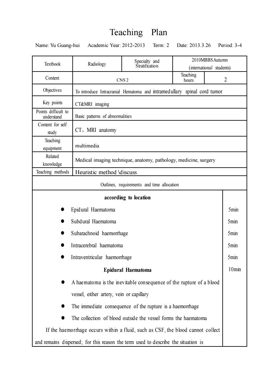
Teaching Plan Name: Yu Guang-hui Academic Year: 2012-2013 Term: 2 Date: 2013.3.26 Period: 3-4 Textbook Radiology Specialty and Stratification 2010MBBSAutumn (international students) Content CNS 2 Teaching hours 2 Objectives To introduce Intracranial Hematoma and intramedullary spinal cord tumor Key points CT&MRI imaging Points difficult to understand Basic patterns of abnormalities Content for self study CT、MRI anatomy Teaching equipment multimedia Related knowledge Medical imaging technique, anatomy, pathology, medicine, surgery Teaching methods Heuristic method \discuss Outlines, requirements and time allocation according to location ⚫ Epidural Haematoma ⚫ Subdural Haematoma ⚫ Subarachnoid haemorrhage ⚫ Intracerebral haematoma ⚫ Intraventricular haemorrhage Epidural Haematoma ⚫ A haematoma is the inevitable consequence of the rupture of a blood vessel, either artery, vein or capillary ⚫ The immediate consequence of the rupture is a haemorrhage ⚫ The collection of blood outside the vessel forms the haematoma If the haemorrhage occurs within a fluid, such as CSF, the blood cannot collect and remains dispersed; for this reason the term used to describe the situation is 5min 5min 5min 5min 5min 10min

subarachnoid haemorrhage,intraventricular haemomhagee 10min CT is the modality of choice Located between the dura and the inner table of the skull Tearing of the meningeal vessels especial middle meningeal artery The shape is usually biconvex ●May cross the mid line Associated with a skull fracture Subdural Haematoma Subdural hematoma is a collection of blood in the subdural space.The main 10min sources of bleeding are thom bridging veins.Rarely a subdural hematoma may be due to rupture of an aneurysm and subsequent laceration of the arachnoidal membraneSubdural hematomas are mainly frontoparietal in location and are usually crescentic in shapeSubdural blood may extend along the falx.in the midline.thus producing interhemispheric subdural hematomas. CT is the examination of choice 5min The density varies according to age of the hematoma:an acute hematoma is hyperdense.becomes isodense in the subacute phase hypodense when chronic(i.e.more than 12 weeks old Space-occupying effect subarachnoid haemorrhage 10min An acute extravasation of blood into the subarachnoid space CT scan clearly shows the hyperdensity of the subarachnoid spaces filled with blood Usually following rupture of an aneurysm of the circle of Willis Blood is cleared from the subarachnoid spaces within 1week Intramedullary spinal cord tumor Intramedullary spinal cord tumor is a tumor that arise from cells of thespinal cord 5min
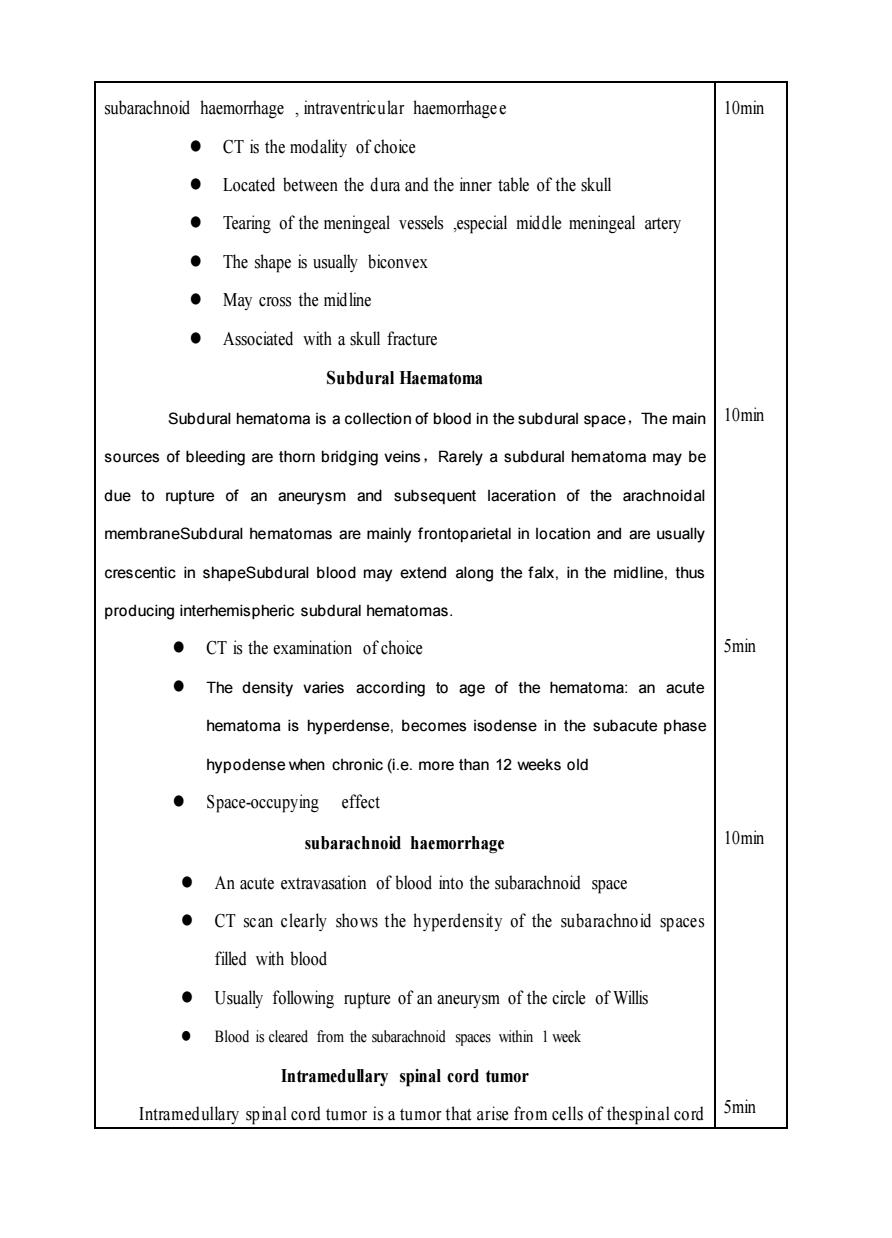
subarachnoid haemorrhage , intraventricular haemorrhage e ⚫ CT is the modality of choice ⚫ Located between the dura and the inner table of the skull ⚫ Tearing of the meningeal vessels ,especial middle meningeal artery ⚫ The shape is usually biconvex ⚫ May cross the midline ⚫ Associated with a skull fracture Subdural Haematoma Subdural hematoma is a collection of blood in the subdural space,The main sources of bleeding are thorn bridging veins,Rarely a subdural hematoma may be due to rupture of an aneurysm and subsequent laceration of the arachnoidal membraneSubdural hematomas are mainly frontoparietal in location and are usually crescentic in shapeSubdural blood may extend along the falx, in the midline, thus producing interhemispheric subdural hematomas. ⚫ CT is the examination of choice ⚫ The density varies according to age of the hematoma: an acute hematoma is hyperdense, becomes isodense in the subacute phase hypodense when chronic (i.e. more than 12 weeks old ⚫ Space-occupying effect subarachnoid haemorrhage ⚫ An acute extravasation of blood into the subarachnoid space ⚫ CT scan clearly shows the hyperdensity of the subarachnoid spaces filled with blood ⚫ Usually following rupture of an aneurysm of the circle of Willis ⚫ Blood is cleared from the subarachnoid spaces within 1 week Intramedullary spinal cord tumor Intramedullary spinal cord tumor is a tumor that arise from cells of thespinal cord 10min 10min 5min 10min 5min

and grow within it The most common histological types are:ependymomas (about 5min 45%)astrocytomas (about 45%),haemangioblastomas (about 5%) and others (about 5%) The clinical presentation is usually slowly progressive 5min Diagnostically plain films of the spine are rarely positive .CT is rarely useful.since the resolution within the spinal canal is 5min limited and only indirect signs of thinning of the pedicles and enlargement of the spinal canal.when present.may be seen MR is the gold standard today 5min
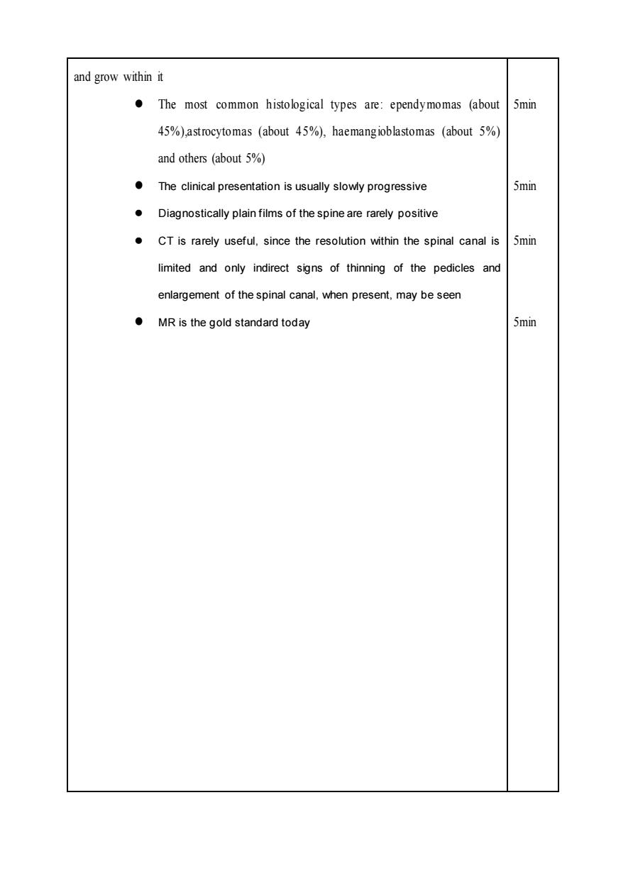
and grow within it ⚫ The most common histological types are: ependymomas (about 45%),astrocytomas (about 45%), haemangioblastomas (about 5%) and others (about 5%) ⚫ The clinical presentation is usually slowly progressive ⚫ Diagnostically plain films of the spine are rarely positive ⚫ CT is rarely useful, since the resolution within the spinal canal is limited and only indirect signs of thinning of the pedicles and enlargement of the spinal canal, when present, may be seen ⚫ MR is the gold standard today 5min 5min 5min 5min