Teaching Plan Name:Linxiang Liu Academic Year:2012-2013 Term:2 Date:May 13 Period:5-6 Textbook Diagnostic Imaging 8e8 2010 MBBS autumn (students) Content Pulmonary Tumors 2 Objectives Pulmonary Tumors Key points Lung cancers Pon市tmao unders Lung cancers Content for self study CT、MR anatomy Teaching equipment multimedia Related knowledge Medical imaging technique,anatomy,pathology,medicine,surgery Teaching methods Heuristic method discuss Outlines,requirements and time allocation Tumors of lungs Benign tumor 10 ● sarco Lung cancers 10 Pthology-Radiology typesquol arcinoma,Adencarcinoma. 15 Central type:direct signs:hilar mass,obturation of bronchus; indirect signs:obstructive emphysema,obstructive atelectasis:transverse S sign, obstructive inflammation,metastatic signs:changes of hilum,changes of eripheral ually 20 of with or with multiple nodules,unilateral or bilateral.pleural metastasis:pleural effusion lymphoadenopathy of hilum.chest wall infiltration destruction of ribs,diaphragm 10 in most c mass.metastatic signs may also show
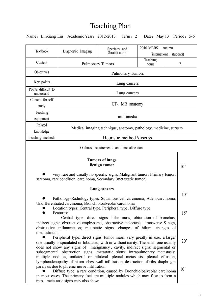
1 Teaching Plan Name:Linxiang Liu Academic Year:2012-2013 Term:2 Date:May 13 Period:5-6 Textbook Diagnostic Imaging Specialty and Stratification 2010 MBBS autumn (international students) Content Pulmonary Tumors Teaching hours 2 Objectives Pulmonary Tumors Key points Lung cancers Points difficult to understand Lung cancers Content for self study CT、MR anatomy Teaching equipment multimedia Related knowledge Medical imaging technique, anatomy, pathology, medicine, surgery Teaching methods Heuristic method \discuss Outlines, requirements and time allocation Tumors of lungs Benign tumor ⚫ very rare and usually no specific signs. Malignant tumor: Primary tumor: sarcoma, rare condition, carcinoma, Secondary (metastatic tumor) Lung cancers ⚫ Pathology-Radiology types: Squamous cell carcinoma, Adenocarcinoma, Undifferentiated carcinoma, Bronchioloalveolar carcinoma ⚫ Location types: Central type, Peripheral type, Diffuse type ⚫ Features: Central type: direct signs: hilar mass, obturation of bronchus; indirect signs: obstructive emphysema, obstructive atelectasis:transverse S sign, obstructive inflammation; metastatic signs: changes of hilum, changes of mediastinum. ⚫ Peripheral type: direct signs: tumor mass: vary greatly in size, a larger one usually is spiculated or lobulated, with or without cavity. The small one usually does not show any signs of malignancy., cavity. indirect signs: segmental or subsegmental obstruction signs. metastatic signs: intrapulmonary metastasis: multiple nodules, unilateral or bilateral. pleural metastasis: pleural effusion, lymphoadenopathy of hilum. chest wall infiltration: destruction of ribs, diaphragm paralysis due to phrenic nerve infiltration. ⚫ Diffuse type: a rare condition, caused by Bronchioloalveolar carcinoma in most cases. The primary foci are multiple nodules which may fuse to form a mass. metastatic signs may also show. 10’ 10’ 15’ 20’ 10’

Pancoast'sneoplasm 15 hand orer's syndrome,destruction of bone,atrophy of C+CT and thin collimation and MRI are the preferred modalities for evaluating Metastatic tumor of lungs 20 The lungs are the common organs of metastasis for most malignancies Lngem is the most important procedure for cancer staging Features:multiple nodules throughout the lungs,usually round and smooth in margin,with or without cavity
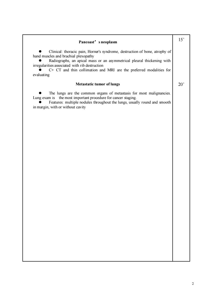
2 Pancoast’s neoplasm ⚫ Clinical: thoracic pain, Horner's syndrome, destruction of bone, atrophy of hand muscles and brachial plexopathy ⚫ Radiographs, an apical mass or an asymmetrical pleural thickening with irregularities associated with rib destruction ⚫ C+ CT and thin collimation and MRI are the preferred modalities for evaluating Metastatic tumor of lungs ⚫ The lungs are the common organs of metastasis for most malignancies. Lung exam is the most important procedure for cancer staging. ⚫ Features: multiple nodules throughout the lungs, usually round and smooth in margin, with or without cavity 15’ 20’

3
3