Teaching Plan Name:BaoJian Wang Academic Year:2012-2013 Term:2 Date.April 9 Period:3-4 2010 MBBS autumr Textbook Diagnosic Imaging Sonnd Content Gastrointestinal (GI)Imaging1 Teaching hours 2 Objectives To grasp the normal appearances of Gl Key points How to read the films of barium examination poat可 Special terms used on barium Content for self study The adverse effect of barium examination Teaching equipment multimedia Related knowedge Physics of X-ray Teaching methods Heuristic method /discuss Outlines requirements and time allocation Anatomy of Gastrointestinal tract Food passes through a long tube inside the body known as the alimentary canal or the gastrointestinal tract(GI tract).The alimentary canal is made up of the oral to digest food but do not have food pass through them.Accessor obnne system include the teeth,tongue,salvary glands,Iive ●Mouth ●Pharynx ●Esophagus ●Stomach ●Small Intestine ●Large Intestine The Imaging modalities 10 ●Radi (X) tinal Tr maging (UGI) Computed Tomography (CT) .Magnetic Resonance Imaging (MRI)
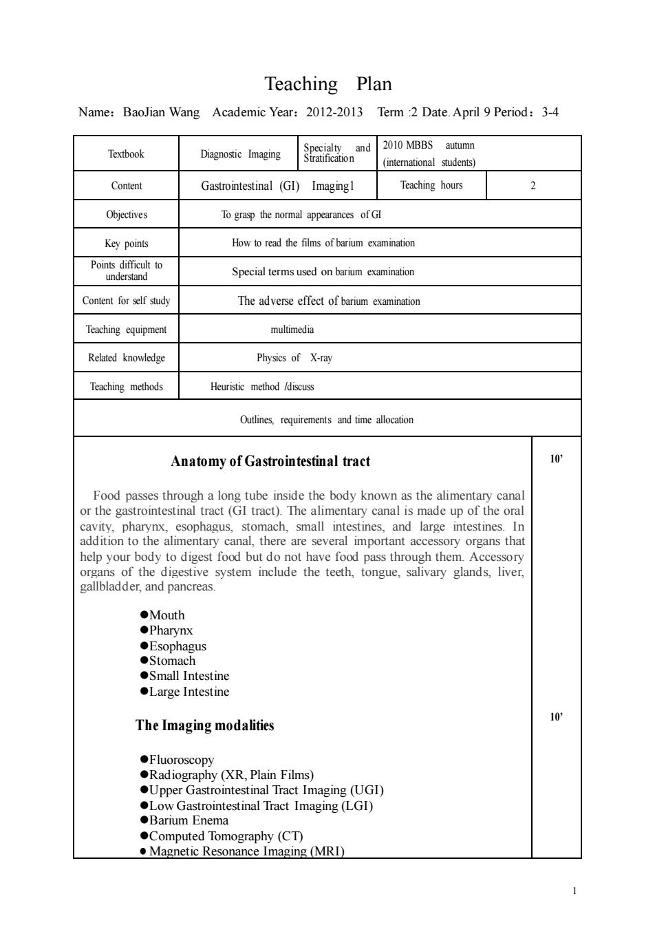
1 Teaching Plan Name:BaoJian Wang Academic Year:2012-2013 Term :2 Date.April 9 Period:3-4 Textbook Diagnostic Imaging Specialty and Stratification 2010 MBBS autumn (international students) Content Gastrointestinal (GI) Imaging1 Teaching hours 2 Objectives To grasp the normal appearances of GI Key points How to read the films of barium examination Points difficult to understand Special terms used on barium examination Content for self study The adverse effect of barium examination Teaching equipment multimedia Related knowledge Physics of X-ray Teaching methods Heuristic method /discuss Outlines, requirements and time allocation Anatomy of Gastrointestinal tract Food passes through a long tube inside the body known as the alimentary canal or the gastrointestinal tract (GI tract). The alimentary canal is made up of the oral cavity, pharynx, esophagus, stomach, small intestines, and large intestines. In addition to the alimentary canal, there are several important accessory organs that help your body to digest food but do not have food pass through them. Accessory organs of the digestive system include the teeth, tongue, salivary glands, liver, gallbladder, and pancreas. ⚫Mouth ⚫Pharynx ⚫Esophagus ⚫Stomach ⚫Small Intestine ⚫Large Intestine The Imaging modalities ⚫Fluoroscopy ⚫Radiography (XR, Plain Films) ⚫Upper Gastrointestinal Tract Imaging (UGI) ⚫Low Gastrointestinal Tract Imaging (LGI) ⚫Barium Enema ⚫Computed Tomography (CT) ⚫ Magnetic Resonance Imaging (MRI) 10’ 10’

Outlines requirements and time allocation X-RAY(Fluoroscopy,Radiography):Normal abdomen. to. AP view of abdomen in supine position ◆Normal bowel gas: Low density. ●Bones::High density All abdominal organs:Water density. UGI(belongs to Barium examination,BE) lo Medical i LGI(Barium Enema) Lower gastrointestinal (GI)tract radiography,also called a lower GI or barium enema,is an x-ray examination of the large intestine,also known as the colon.This examination evaluates the right or ascending colon,the transverse colon,the left or Abdominal CT scan 10 CT allows high-quality multiplanar reformation and three-dimensional reconstruction of gastric images. CT provides information about both the gastric wall and the extragastric extent of disease.Preoperative staging of gastric carcinoma appears to be the main clinical indication for multidetector CT.Multidetector CT is a valuable tool for the evaluation of gastric wall disease and serves as an adjunct to endoscopy. Abdominal MR scan 10 An MRI of the Abdomen obtains high-resolution images of the contents of the abdomen,including the liver,spleen,adrenal glands,pancreas,kidneys,G.I.Tract, and supporting mesentery.It is usually obtained with and without contrast.(IV required) Operation Positions of Barium Examination lo. ●Erect, ●supine,. ●prone left decubitus
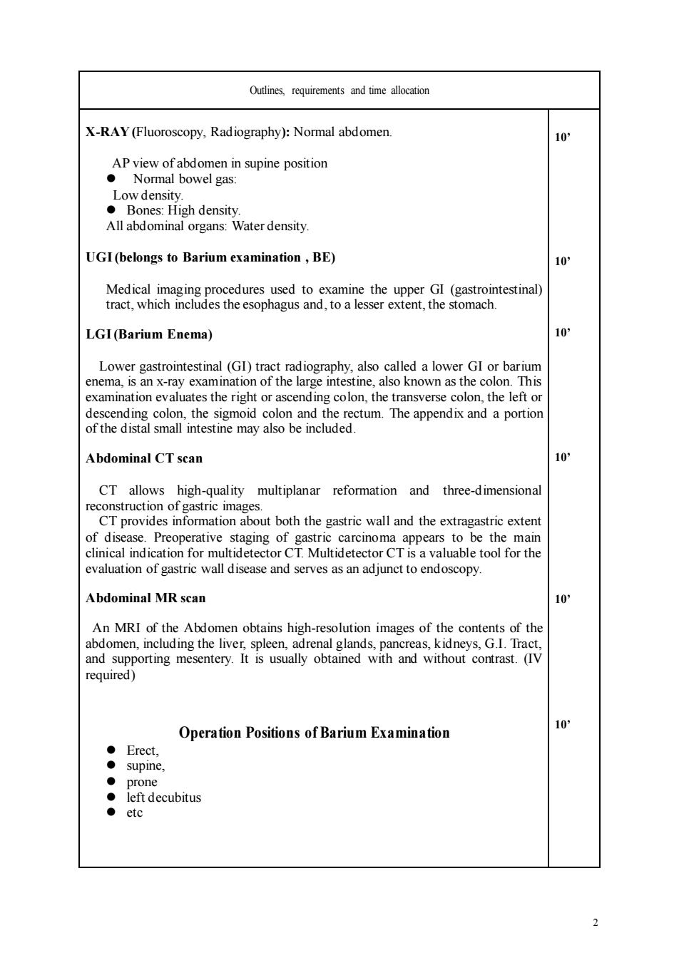
2 Outlines, requirements and time allocation X-RAY (Fluoroscopy, Radiography): Normal abdomen. AP view of abdomen in supine position ⚫ Normal bowel gas: Low density. ⚫ Bones: High density. All abdominal organs: Water density. UGI (belongs to Barium examination , BE) Medical imaging procedures used to examine the upper GI (gastrointestinal) tract, which includes the esophagus and, to a lesser extent, the stomach. LGI (Barium Enema) Lower gastrointestinal (GI) tract radiography, also called a lower GI or barium enema, is an x-ray examination of the large intestine, also known as the colon. This examination evaluates the right or ascending colon, the transverse colon, the left or descending colon, the sigmoid colon and the rectum. The appendix and a portion of the distal small intestine may also be included. Abdominal CT scan CT allows high-quality multiplanar reformation and three-dimensional reconstruction of gastric images. CT provides information about both the gastric wall and the extragastric extent of disease. Preoperative staging of gastric carcinoma appears to be the main clinical indication for multidetector CT. Multidetector CT is a valuable tool for the evaluation of gastric wall disease and serves as an adjunct to endoscopy. Abdominal MR scan An MRI of the Abdomen obtains high-resolution images of the contents of the abdomen, including the liver, spleen, adrenal glands, pancreas, kidneys, G.I. Tract, and supporting mesentery. It is usually obtained with and without contrast. (IV required) Operation Positions of Barium Examination ⚫ Erect, ⚫ supine, ⚫ prone ⚫ left decubitus ⚫ etc 10’ 10’ 10’ 10’ 10’ 10’

Outlines,requirements and time allocation Principles of Image Analysis Appearance of the Stomach 10* The radiologist first examines the overall position,shape,and size of the stomach.The gastric fundusabuts the left hemidiaphragm.The cardia hasa midline location,abuttingthe crus ofthe left hemidiaphragm. The stomach curves to the right across the midline,with the distal gastric antrum and duodenum extendingto the right of the spine.There is considerable variation in the size of the stomach,dependingon the amount of barium and effervescent agent administered. Luminal Contour 5 In the barium pool,the contour is demarcated by a smooth edge of barium.With air contrast. the luminal contourappearsasa smooth,continuous barium-coated white line. Barium Pool The pool of high-density barium is the tool the radiologist uses to scrub and coat the mucosal surface. 3
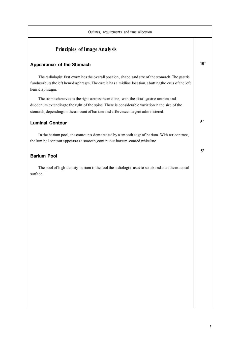
3 Outlines, requirements and time allocation Principles of Image Analysis Appearance of the Stomach The radiologist first examines the overall position, shape, and size of the stomach. The gastric fundus abuts the left hemidiaphragm. The cardia has a midline location, abutting the crus of the left hemidiaphragm. The stomach curves to the right across the midline, with the distal gastric antrum and duodenum extending to the right of the spine. There is considerable variation in the size of the stomach, depending on the amount of barium and effervescent agent administered. Luminal Contour In the barium pool, the contour is demarcated by a smooth edge of barium. With air contrast, the luminal contour appears as a smooth, continuous barium-coated white line. Barium Pool The pool of high-density barium is the tool the radiologist uses to scrub and coat the mucosal surface. 10’ 5’ 5’