正在加载图片...
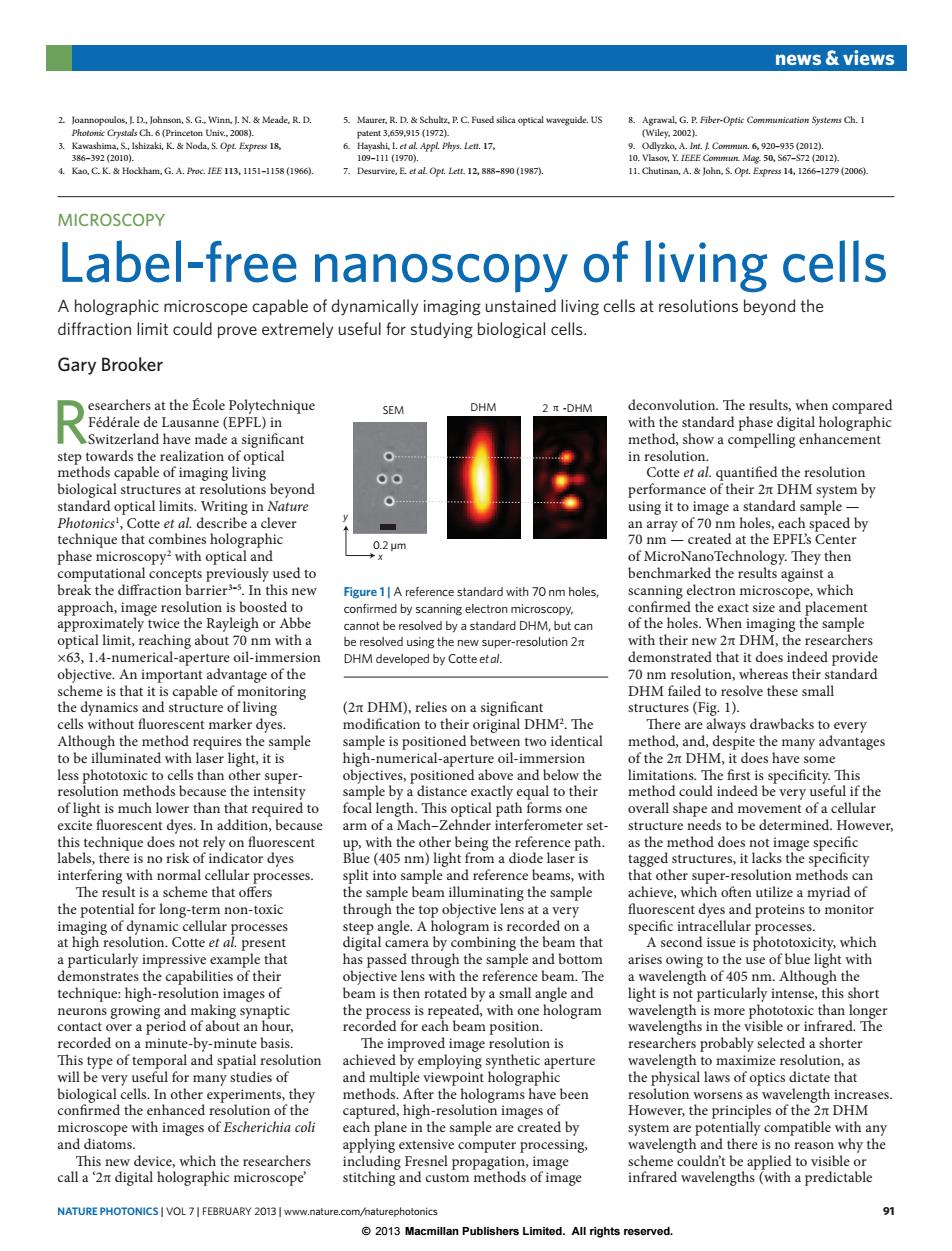
news views 2.Joannopoulos.ID.Johnson.S.G.Winn.I.N.&Meade.R.D. 5.Maurer.R.D.Schultz,P.C.Fused silica optical waveguide.US 8.Agrawal,G.P.Fiber-Optic Communication Systems Ch.I Photomic Crystals Ch.6(Princeton Univ..2008). patent3,659,915(1972. (Wiley.2002). 3.Kawashima,S.,Ishizaki,K.Noda.S.Opt.Express 18. 6.Hayashi,1.et al.Appl.Phys.Lett.17, 9.Odlyzko,A.m.1 Commun.6,920-935(2012. 386-39220101. 109-11141970). 10.Vlasov,Y.IEEE Common Mag.50,S67-S72 (2012). 4.K0,C.K.&Hockham,G.A.Proc.IEE113,1151-158(1966). 7 Desurvire,E.et al Opt.Lett.12,888-890(1987). 11.Chutinan,A.John.S.Opt.Express 14,1266-1279 (2006). MICROSCOPY Label-free nanoscopy of living cells A holographic microscope capable of dynamically imaging unstained living cells at resolutions beyond the diffraction limit could prove extremely useful for studying biological cells. Gary Brooker esearchers at the Ecole Polytechnique SEM DHM 2π-DHM deconvolution.The results,when compared Federale de Lausanne(EPFL)in with the standard phase digital holographic Switzerland have made a significant method,show a compelling enhancement step towards the realization of optical in resolution. methods capable of imaging living Cotte et al.quantified the resolution ● biological structures at resolutions beyond performance of their 2t DHM system by standard optical limits.Writing in Nature using it to image a standard sample- Photonics',Cotte et al.describe a clever an array of 70 nm holes,each spaced by technique that combines holographic 0.2μm 70 nm-created at the EPFL's Center phase microscopy:with optical and of MicroNanoTechnology.They then computational concepts previously used to benchmarked the results against a break the diffraction barrier3-5.In this new Figure 1|A reference standard with 70 nm holes, scanning electron microscope,which approach,image resolution is boosted to confirmed by scanning electron microscopy, confirmed the exact size and placement approximately twice the Rayleigh or Abbe cannot be resolved by a standard DHM,but can of the holes.When imaging the sample optical limit,reaching about 70 nm with a be resolved using the new super-resolution 2n with their new 2nt DHM,the researchers x63,1.4-numerical-aperture oil-immersion DHM developed by Cotte etal. demonstrated that it does indeed provide objective.An important advantage of the 70 nm resolution,whereas their standard scheme is that it is capable of monitoring DHM failed to resolve these small the dynamics and structure of living (2n DHM),relies on a significant structures (Fig.1). cells without fluorescent marker dyes. modification to their original DHM2.The There are always drawbacks to every Although the method requires the sample sample is positioned between two identical method,and,despite the many advantages to be illuminated with laser light,it is high-numerical-aperture oil-immersion of the 2nt DHM,it does have some less phototoxic to cells than other super- objectives,positioned above and below the limitations.The first is specificity.This resolution methods because the intensity sample by a distance exactly equal to their method could indeed be very useful if the of light is much lower than that required to focal length.This optical path forms one overall shape and movement of a cellular excite fluorescent dyes.In addition,because arm of a Mach-Zehnder interferometer set- structure needs to be determined.However this technique does not rely on fluorescent up,with the other being the reference path. as the method does not image specific labels,there is no risk of indicator dyes Blue (405 nm)light from a diode laser is tagged structures,it lacks the specificity interfering with normal cellular processes. split into sample and reference beams,with that other super-resolution methods can The result is a scheme that offers the sample beam illuminating the sample achieve,which often utilize a myriad of the potential for long-term non-toxic through the top objective lens at a very fluorescent dyes and proteins to monitor imaging of dynamic cellular processes steep angle.A hologram is recorded on a specific intracellular processes. at high resolution.Cotte et al.present digital camera by combining the beam that A second issue is phototoxicity,which a particularly impressive example that has passed through the sample and bottom arises owing to the use of blue light with demonstrates the capabilities of their objective lens with the reference beam.The a wavelength of 405 nm.Although the technique:high-resolution images of beam is then rotated by a small angle and light is not particularly intense,this short neurons growing and making synaptic the process is repeated,with one hologram wavelength is more phototoxic than longer contact over a period of about an hour, recorded for each beam position. wavelengths in the visible or infrared.The recorded on a minute-by-minute basis. The improved image resolution is researchers probably selected a shorter This type of temporal and spatial resolution achieved by employing synthetic aperture wavelength to maximize resolution,as will be very useful for many studies of and multiple viewpoint holographic the physical laws of optics dictate that biological cells.In other experiments,they methods.After the holograms have been resolution worsens as wavelength increases. confirmed the enhanced resolution of the captured,high-resolution images of However,the principles of the 2nt DHM microscope with images of Escherichia coli each plane in the sample are created by system are potentially compatible with any and diatoms. applying extensive computer processing, wavelength and there is no reason why the This new device,which the researchers including Fresnel propagation,image scheme couldn't be applied to visible or call a '2n digital holographic microscope stitching and custom methods of image infrared wavelengths(with a predictable NATURE PHOTONICS VOL 7|FEBRUARY 2013 www.nature.com/naturephotonics 91 2013 Macmillan Publishers Limited.All rights reserved© 2013 Macmillan Publishers Limited. All rights reserved. NATURE PHOTONICS | VOL 7 | FEBRUARY 2013 | www.nature.com/naturephotonics 91 news & views 2. Joannopoulos, J. D., Johnson, S. G., Winn, J. N. & Meade, R. D. Photonic Crystals Ch. 6 (Princeton Univ., 2008). 3. Kawashima, S., Ishizaki, K. & Noda, S. Opt. Express 18, 386–392 (2010). 4. Kao, C. K. & Hockham, G. A. Proc. IEE 113, 1151–1158 (1966). 5. Maurer, R. D. & Schultz, P. C. Fused silica optical waveguide. US patent 3,659,915 (1972). 6. Hayashi, I. et al. Appl. Phys. Lett. 17, 109–111 (1970). 7. Desurvire, E. et al. Opt. Lett. 12, 888–890 (1987). 8. Agrawal, G. P. Fiber-Optic Communication Systems Ch. 1 (Wiley, 2002). 9. Odlyzko, A. Int. J. Commun. 6, 920–935 (2012). 10. Vlasov, Y. IEEE Commun. Mag. 50, S67–S72 (2012). 11. Chutinan, A. & John, S. Opt. Express 14, 1266–1279 (2006). Researchers at the École Polytechnique Fédérale de Lausanne (EPFL) in Switzerland have made a significant step towards the realization of optical methods capable of imaging living biological structures at resolutions beyond standard optical limits. Writing in Nature Photonics1 , Cotte et al. describe a clever technique that combines holographic phase microscopy2 with optical and computational concepts previously used to break the diffraction barrier3–5. In this new approach, image resolution is boosted to approximately twice the Rayleigh or Abbe optical limit, reaching about 70 nm with a ×63, 1.4-numerical-aperture oil-immersion objective. An important advantage of the scheme is that it is capable of monitoring the dynamics and structure of living cells without fluorescent marker dyes. Although the method requires the sample to be illuminated with laser light, it is less phototoxic to cells than other superresolution methods because the intensity of light is much lower than that required to excite fluorescent dyes. In addition, because this technique does not rely on fluorescent labels, there is no risk of indicator dyes interfering with normal cellular processes. The result is a scheme that offers the potential for long-term non-toxic imaging of dynamic cellular processes at high resolution. Cotte et al. present a particularly impressive example that demonstrates the capabilities of their technique: high-resolution images of neurons growing and making synaptic contact over a period of about an hour, recorded on a minute-by-minute basis. This type of temporal and spatial resolution will be very useful for many studies of biological cells. In other experiments, they confirmed the enhanced resolution of the microscope with images of Escherichia coli and diatoms. This new device, which the researchers call a ‘2π digital holographic microscope’ (2π DHM), relies on a significant modification to their original DHM2 . The sample is positioned between two identical high-numerical-aperture oil-immersion objectives, positioned above and below the sample by a distance exactly equal to their focal length. This optical path forms one arm of a Mach–Zehnder interferometer setup, with the other being the reference path. Blue (405 nm) light from a diode laser is split into sample and reference beams, with the sample beam illuminating the sample through the top objective lens at a very steep angle. A hologram is recorded on a digital camera by combining the beam that has passed through the sample and bottom objective lens with the reference beam. The beam is then rotated by a small angle and the process is repeated, with one hologram recorded for each beam position. The improved image resolution is achieved by employing synthetic aperture and multiple viewpoint holographic methods. After the holograms have been captured, high-resolution images of each plane in the sample are created by applying extensive computer processing, including Fresnel propagation, image stitching and custom methods of image deconvolution. The results, when compared with the standard phase digital holographic method, show a compelling enhancement in resolution. Cotte et al. quantified the resolution performance of their 2π DHM system by using it to image a standard sample — an array of 70 nm holes, each spaced by 70 nm — created at the EPFL’s Center of MicroNanoTechnology. They then benchmarked the results against a scanning electron microscope, which confirmed the exact size and placement of the holes. When imaging the sample with their new 2π DHM, the researchers demonstrated that it does indeed provide 70 nm resolution, whereas their standard DHM failed to resolve these small structures (Fig. 1). There are always drawbacks to every method, and, despite the many advantages of the 2π DHM, it does have some limitations. The first is specificity. This method could indeed be very useful if the overall shape and movement of a cellular structure needs to be determined. However, as the method does not image specific tagged structures, it lacks the specificity that other super-resolution methods can achieve, which often utilize a myriad of fluorescent dyes and proteins to monitor specific intracellular processes. A second issue is phototoxicity, which arises owing to the use of blue light with a wavelength of 405 nm. Although the light is not particularly intense, this short wavelength is more phototoxic than longer wavelengths in the visible or infrared. The researchers probably selected a shorter wavelength to maximize resolution, as the physical laws of optics dictate that resolution worsens as wavelength increases. However, the principles of the 2π DHM system are potentially compatible with any wavelength and there is no reason why the scheme couldn’t be applied to visible or infrared wavelengths (with a predictable MICROSCOPY Label-free nanoscopy of living cells A holographic microscope capable of dynamically imaging unstained living cells at resolutions beyond the diffraction limit could prove extremely useful for studying biological cells. Gary Brooker SEM DHM 2 π -DHM 0.2 µm x y Figure 1 | A reference standard with 70 nm holes, confirmed by scanning electron microscopy, cannot be resolved by a standard DHM, but can be resolved using the new super-resolution 2π DHM developed by Cotte et al