正在加载图片...

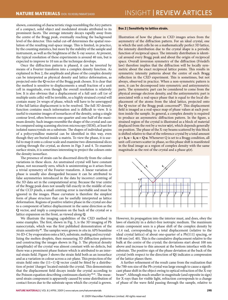
NATURE MATERIALS DOL:10.1038/NMAT2400 INSIGHT I REVIEW ARTICLES shown,consisting of characteristic rings resembling the Airy pattern of a compact,solid object and modulated streaks attributed to its Box 2 Sensitivity to lattice strain. prominent facets.The average intensity decays rapidly away from the centre of the Bragg peak,eventually reaching the background Illustration of how the phase in CXD images arises from the level of the detector.This radial cut-off determines the spatial reso- asymmetry of the diffraction pattern.For an ideal crystal,one lution of the resulting real-space image.This is limited,in practice, in which the unit cells lie on a mathematically perfect 3D lattice, by the counting statistics,but more by the stability of the sample and the intensity distribution due to the crystal shape is a periodic instrument,as well as the brightness of the X-ray source.At present, function of reciprocal space.The intensity distribution is identi- the typical resolution of these experiments is around 40 nm,but is cal around every Bragg peak and about the origin of reciprocal expected to improve to 10 nm as the technique develops. space.Overall inversion symmetry of the diffraction(Friedel's Once the diffraction pattern is phased,it can be inverted by law)therefore implies that the diffraction will be locally sym- means of a Fourier transform into a complex density function.As metric about the exact reciprocal lattice points.This results in explained in Box 2,the amplitude and phase of the complex density symmetric intensity patterns about the centre of each Bragg can be interpreted as physical density and lattice deformation,as reflection in the CXD experiment.This is sometimes,but not projected onto the Q vector of the Bragg peak chosen.It is clear that always,observed in practice.When a non-symmetric pattern is the method is sensitive to displacements a small fraction of a unit seen,it can be decomposed into symmetric and antisymmetric cell in magnitude,even though the overall resolution is relatively parts.The symmetric part can be considered to come from the low.It is also obvious that a displacement of a full unit cell (or of physical average electron density,and the antisymmetric part is multiple units cells)will be invisible,so a highly strained crystal will associated with a real-space phase that is equal to the local dis- contain many 2nt wraps of phase,which will have to be unwrapped placement of the atoms from the ideal lattice,projected onto if the full lattice displacement is to be resolved.The full 3D density the Q vector of the Bragg peak concerned 2.This displacement function contains much information and is difficult to view.It is field is imaged as a real-space map of phase values at each posi- useful to examine images of isosurfaces of the density,using a single tion inside the sample.In general,a complex density is required contour level,often between one-quarter and one-half of the maxi- to produce an asymmetric diffraction pattern.In the figure,a mum density.Such images resemble the shape of the crystal and can strained region of the crystal is illustrated as a block of material be compared using scanning electron microscopy(SEM),at least for displaced from the rest by a vector u(r),which depends generally isolated nanocrystals on a substrate.The shapes of individual grains on position.The phase of the X-ray beams scattered by this block of a polycrystalline material can be identified in this way,even is shifted relative to that of the reference crystal by a total amount though they are buried inside a matrix.To view the phase,it is use- =keu-k-u=Q-u.Whenever Q is set to a Bragg condition,all ful to map it onto a colour scale and examine two-dimensional slices unit-cell corners scatter in phase,so this phase shift is manifested cutting through the crystal,as shown in Figs 3 and 4.To examine in the final image as a region of complex density with the same surface strain,it is sometimes interesting to project the colours onto magnitude as the rest of the crystal and a phase o(r). the density isosurface. The presence of strain can be discerned directly from the colour variations in these slices.An unstrained crystal will have constant phase,not necessarily zero,which is uninteresting as it arises from a trivial symmetry of the Fourier transform.A linear variation of phase is usually also disregarded because it can be attributed to false asymmetries introduced in the data by incorrect centring of the 3D data set in the computational array.Because the true centre of the Bragg peak does not usually fall exactly in the middle of one of the CCD pixels,a small centring error is inevitable and must be ignored in the images.Phase curvature is therefore the simplest form of phase structure that can be usefully interpreted as lattice deformation.Regions of positive relative phase in the crystal are due to a component of lattice displacement in the same direction as the Q vector,and imply a compression on the back of the crystal or a lattice expansion on the front,as viewed along Q. We illustrate the imaging capabilities of the CXD method in However,its propagation into the interior must,and does,obey the some examples.The first,shown in Fig.3,is the 3D imaging of Pb laws of elasticity in a defect-free isotropic medium.The maximum nanocrystals,which was the first published demonstration of the strain component seen is a phase shift of the complex density by strain sensitivity.The samples were grown in situ in APS beamline +1.4 rad,corresponding to a total displacement (relative to the 34-ID-Cby evaporation onto a SiO,substrate,melting and recrystal- ideal crystal lattice)of about one-quarter of a Pb{111}spacing,or lizing the molten droplets,then measuring 3D diffraction patterns 0.08 nm (ref.40).This is the cumulative displacement relative to the and constructing the images shown in Fig.3.The physical density bulk at the centre of the crystal;the deviations start about 100 nm (amplitude)of the crystal was almost constant with no defects,but above and increase to this amount at the bottom interface with the there was a prominent phase feature which is attributed to an inter- substrate.The positive sign of the phase deviation at the back of the nal strain field.Figure 3 shows the strain field both as an isosurface crystal (with respect to the direction of Q)indicates a compression and as a variation in colour across a cut-plane.This projection of the of the lattice planes there. strain field onto the (11-1)Q vector could be fitted by a distribu- A further refinement of the result came from the realization that tion of point 'charges'located outside the crystal.This demonstrates the 700-nm size of the Pb crystal was large enough to cause a signifi- that the displacement field decays inside the crystal according to cant phase shift in the object owing to optical refraction of the X-ray the Poisson equation describing continuum elasticity354.The meas- beam.Although much smaller in magnitude(and opposite in sign) ured strain component is apparently caused by some distribution of for X-rays than for visible light,refraction corresponds to a change contact forces due to the substrate upon which the crystal is grown. of phase of the wave field passing through the sample,relative to NATURE MATERIALS VOL 8|APRIL 2009 www.nature.com/naturematerials 295 2009 Macmillan Publishers Limited.All rights reservednature materials | VOL 8 | APRIL 2009 | www.nature.com/naturematerials 295 NaTure maTerials doi: 10.1038/nmat2400 insight | review articles However, its propagation into the interior must, and does, obey the laws of elasticity in a defect-free isotropic medium. The maximum strain component seen is a phase shift of the complex density by +1.4 rad, corresponding to a total displacement (relative to the ideal crystal lattice) of about one-quarter of a Pb{111} spacing, or 0.08 nm (ref. 40). This is the cumulative displacement relative to the bulk at the centre of the crystal; the deviations start about 100 nm above and increase to this amount at the bottom interface with the substrate. The positive sign of the phase deviation at the back of the crystal (with respect to the direction of Q) indicates a compression of the lattice planes there. A further refinement of the result came from the realization that the 700-nm size of the Pb crystal was large enough to cause a significant phase shift in the object owing to optical refraction of the X-ray beam42. Although much smaller in magnitude (and opposite in sign) for X-rays than for visible light, refraction corresponds to a change of phase of the wave field passing through the sample, relative to shown, consisting of characteristic rings resembling the Airy pattern of a compact, solid object and modulated streaks attributed to its prominent facets. The average intensity decays rapidly away from the centre of the Bragg peak, eventually reaching the background level of the detector. This radial cut-off determines the spatial resolution of the resulting real-space image. This is limited, in practice, by the counting statistics, but more by the stability of the sample and instrument, as well as the brightness of the X-ray source. At present, the typical resolution of these experiments is around 40 nm, but is expected to improve to 10 nm as the technique develops. Once the diffraction pattern is phased, it can be inverted by means of a Fourier transform into a complex density function. As explained in Box 2, the amplitude and phase of the complex density can be interpreted as physical density and lattice deformation, as projected onto the Q vector of the Bragg peak chosen. It is clear that the method is sensitive to displacements a small fraction of a unit cell in magnitude, even though the overall resolution is relatively low. It is also obvious that a displacement of a full unit cell (or of multiple units cells) will be invisible, so a highly strained crystal will contain many 2π wraps of phase, which will have to be unwrapped if the full lattice displacement is to be resolved. The full 3D density function contains much information and is difficult to view. It is useful to examine images of isosurfaces of the density, using a single contour level, often between one-quarter and one-half of the maximum density. Such images resemble the shape of the crystal and can be compared using scanning electron microscopy (SEM), at least for isolated nanocrystals on a substrate. The shapes of individual grains of a polycrystalline material can be identified in this way, even though they are buried inside a matrix. To view the phase, it is useful to map it onto a colour scale and examine two-dimensional slices cutting through the crystal, as shown in Figs 3 and 4. To examine surface strain, it is sometimes interesting to project the colours onto the density isosurface. The presence of strain can be discerned directly from the colour variations in these slices. An unstrained crystal will have constant phase, not necessarily zero, which is uninteresting as it arises from a trivial symmetry of the Fourier transform. A linear variation of phase is usually also disregarded because it can be attributed to false asymmetries introduced in the data by incorrect centring of the 3D data set in the computational array. Because the true centre of the Bragg peak does not usually fall exactly in the middle of one of the CCD pixels, a small centring error is inevitable and must be ignored in the images. Phase curvature is therefore the simplest form of phase structure that can be usefully interpreted as lattice deformation. Regions of positive relative phase in the crystal are due to a component of lattice displacement in the same direction as the Q vector, and imply a compression on the back of the crystal or a lattice expansion on the front, as viewed along Q. We illustrate the imaging capabilities of the CXD method in some examples. The first, shown in Fig. 3, is the 3D imaging of Pb nanocrystals, which was the first published demonstration of the strain sensitivity40. The samples were grown in situ in APS beamline 34-ID-C by evaporation onto a SiO2 substrate, melting and recrystallizing the molten droplets, then measuring 3D diffraction patterns and constructing the images shown in Fig. 3. The physical density (amplitude) of the crystal was almost constant with no defects, but there was a prominent phase feature which is attributed to an internal strain field. Figure 3 shows the strain field both as an isosurface and as a variation in colour across a cut-plane. This projection of the strain field onto the (11−1) Q vector could be fitted by a distribution of point ‘charges’ located outside the crystal. This demonstrates that the displacement field decays inside the crystal according to the Poisson equation describing continuum elasticity35,41. The measured strain component is apparently caused by some distribution of contact forces due to the substrate upon which the crystal is grown. Illustration of how the phase in CXD images arises from the asymmetry of the diffraction pattern. For an ideal crystal, one in which the unit cells lie on a mathematically perfect 3D lattice, the intensity distribution due to the crystal shape is a periodic function of reciprocal space. The intensity distribution is identical around every Bragg peak and about the origin of reciprocal space. Overall inversion symmetry of the diffraction (Friedel’s law) therefore implies that the diffraction will be locally symmetric about the exact reciprocal lattice points. This results in symmetric intensity patterns about the centre of each Bragg reflection in the CXD experiment. This is sometimes, but not always, observed in practice. When a non-symmetric pattern is seen, it can be decomposed into symmetric and antisymmetric parts. The symmetric part can be considered to come from the physical average electron density, and the antisymmetric part is associated with a real-space phase that is equal to the local displacement of the atoms from the ideal lattice, projected onto the Q vector of the Bragg peak concerned62. This displacement field is imaged as a real-space map of phase values at each position inside the sample. In general, a complex density is required to produce an asymmetric diffraction pattern. In the figure, a strained region of the crystal is illustrated as a block of material displaced from the rest by a vector u(r), which depends generally on position. The phase of the X-ray beams scattered by this block is shifted relative to that of the reference crystal by a total amount φ = kf ∙u − ki ∙u = Q∙u. Whenever Q is set to a Bragg condition, all unit-cell corners scatter in phase, so this phase shift is manifested in the final image as a region of complex density with the same magnitude as the rest of the crystal and a phase φ(r). Box 2 | sensitivity to lattice strain. u ki kf nmat_2400_APR09.indd 295 13/3/09 12:04:31 © 2009 Macmillan Publishers Limited. All rights reserved