正在加载图片...
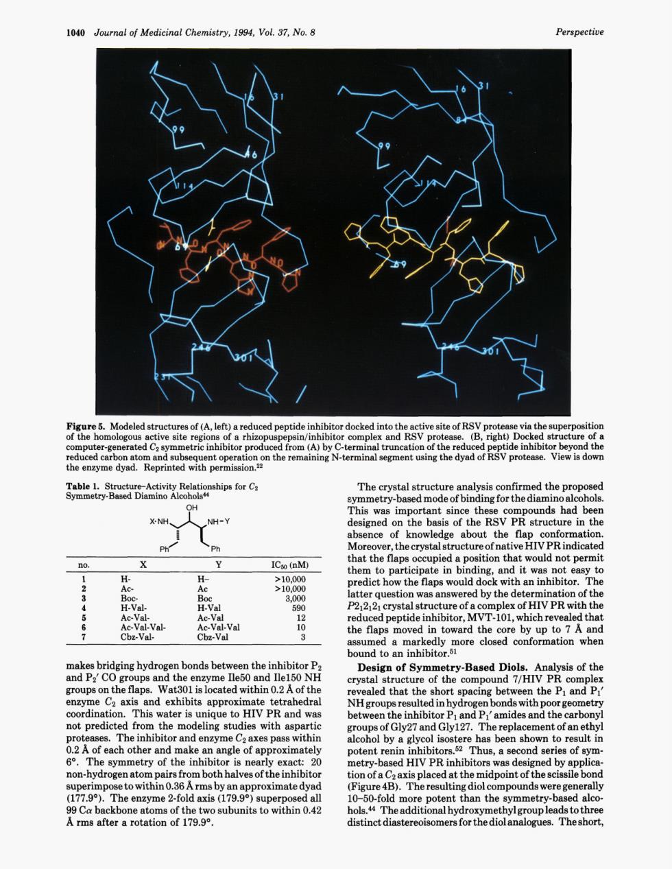
1040 Journal of Medicinal Chemistry,1994,Vol.37,No.8 Perspectiu Table.cActivity Relationhips for C (NH. NH-Y Mor ce ICso (nM) that the CeVa-Val- 20 epeptidemhihior,NTioi,hicdhreregeg d tha bound to an of the ater isunique aed aled that the This w to HIV PR and wa the inhibitor Pand Pamides and the carbo ot pr hus,a second series of sym non-hydrogen ato e 2-fold axis( 上1040 Journal of Medicinal Chemistry, 1994, Vol. 37, No. 8 Perspective Figure 5. Modeled structures of (A, left) a reduced peptide inhibitor docked into the active site of RSV protease via the superposition of the homologous active site regions of a rhizopuspepsin/inhibitor complex and RSV protease. (B, right) Docked structure of a computer-generated CZ symmetric inhibitor produced from (A) by C-terminal truncation of the reduced peptide inhibitor beyond the reduced carbon atom and subsequent operation on the remaining N-terminal segment using the dyad of RSV protease. View is dow the enzyme dyad. Reprinted with permission.22 Table 1. Structure-Activity Relationships for CZ Symmetry-Based Diamino Alcoholsu Ph’ Ph 1 H- H- >lO,OOo 2 AC- Ac >10,000 3 BOC- Boc 3,000 4 H-Val- H-Val 590 5 Ac-Val- Ac-Val 12 6 Ac-Val- Val- Ac-Val-Val 10 7 Cbz-Val- Cbz-Val 3 makes bridging hydrogen bonds between the inhibitor P2 and P2’ CO groups and the enzyme Ile50 and Ile150 NH groups on the flaps. Wat301 is located within 0.2 A of the enzyme C2 axis and exhibits approximate tetrahedral coordination. This water is unique to HIV PR and was not predicted from the modeling studies with aspartic proteases. The inhibitor and enzyme C2 axes pass within 0.2 A of each other and make an angle of approximately 6’. The symmetry of the inhibitor is nearly exact: 20 non-hydrogen atom pairs from both halves of the inhibitor superimpose to within 0.36 Arms by an approximate dyad (177.9’). The enzyme 2-fold axis (179.9O) superposed all 99 Ca backbone atoms of the two subunits to within 0.42 A rms after a rotation of 179.9O. The crystal structure analysis confirmed the proposed symmetry-based mode of binding for the diamino alcohols. This was important since these compounds had been designed on the basis of the RSV PR structure in the absence of knowledge about the flap conformation. Moreover, the crystal structure of native HIV PR indicated that the flaps occupied a position that would not permit them to participate in binding, and it was not easy to predict how the flaps would dock with an inhibitor. The latter question was answered by the determination of the E212121 crystal structure of a complex of HIV PR with the reduced peptide inhibitor, MVT-101, which revealed that the flaps moved in toward the core by up to 7 A and assumed a markedly more closed conformation when bound to an inhibit~r.~~ Design of Symmetry-Based Diols. Analysis of the crystal structure of the compound 7/HIV PR complex revealed that the short spacing between the P1 and PI’ NH groups resulted in hydrogen bonds with poor geometry between the inhibitor P1 and PI’ amides and the carbonyl groups of Gly27 and Gly127. The replacement of an ethyl alcohol by a glycol isostere has been shown to result in potent renin inhibit0rs.5~ Thus, a second series of symmetry-based HIV PR inhibitors was designed by application of a C2 axis placed at the midpoint of the scissile bond (Figure 4B). The resulting diol compounds were generally 10-50-fold more potent than the symmetry-based alcohols.& The additional hydroxymethyl group leads to three distinct diastereoisomers for the diol analogues. The short