正在加载图片...
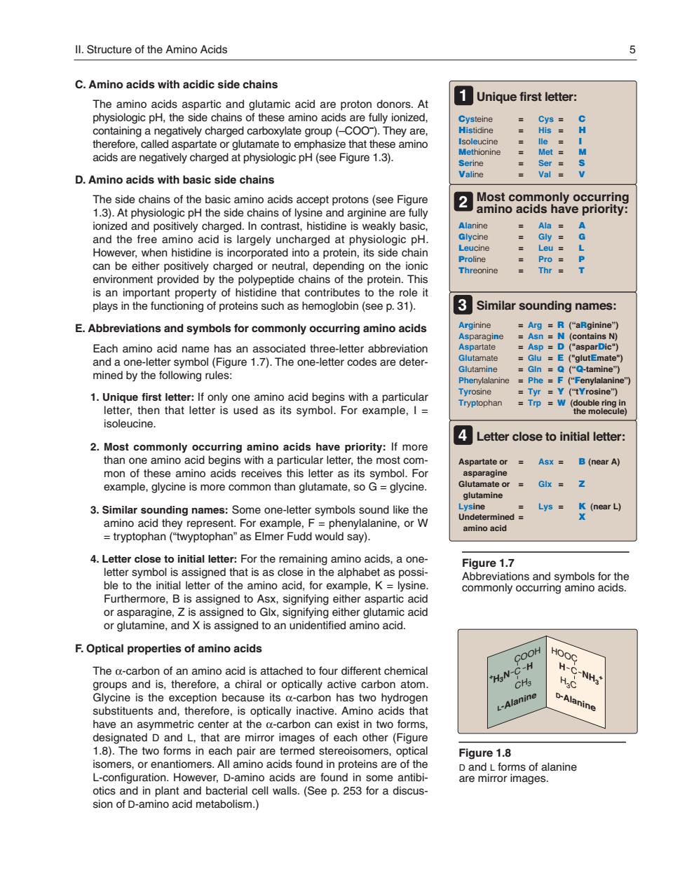
Il.Structure of the Amino Acids C.Amino acids with acidic side chains The amino acids aspartic and glutamic acid are proton donors.At 1Unique first letter: ate or alutamate to e asize that these acids are negatively charged at physiologic pH(see Figure 1.3). : D.Amino acids with basic side chains 13.At h ionized and positively charged.In contrast,histidine is weakly basic idne is incorporated environment provided by the polypeptide chans of the protein.This hreonine is an important property of histidine that contributes to the role it plays in the functioning of proteins such as hemoglobin (see p.31). 3Similar sounding names: E.Abbreviations and symbols for commonly occurring amino acids ArgR (aRginine (e Each aminc acid name has =D and a one-letter mined by the following rules: vr Y (tYrosine" 4Letter close to initial letter: 2 Most co mino Asx B(near A) mon of these amino acids receives this letter as its symbol.For example,glycine is more common than glutamate,so G glycine Lys K( d amino acid 4.Letter close to initial letter:For the remaining amino acids a one letter symbol is assigned that is as close in the alphabet as possi Figure 1.7 ble to the initia terof the amino acid.for example,K-lysine Furthermore, assigne amacd or glutamine.and is assigned to an unidentified amino acid. F.Optical properties of amino acids The a-carbon of an amino acid is attached to four different chemica H has two hydog L-Alanine have an asyr metric center at the a-caon can designated D and L,that are mirror images of each other(Figure Figure1.8 omers. is a otics and in plant and bacterial cell walls.(See p.253 fora discus- 8aeatromsgdsane sion of D-amino acid metabolism.)C. Amino acids with acidic side chains The amino acids aspartic and glutamic acid are proton donors. At physiologic pH, the side chains of these amino acids are fully ionized, containing a negatively charged carboxylate group (–COO– ). They are, therefore, called aspartate or glutamate to emphasize that these amino acids are negatively charged at physiologic pH (see Figure 1.3). D. Amino acids with basic side chains The side chains of the basic amino acids accept protons (see Figure 1.3). At physiologic pH the side chains of lysine and arginine are fully ionized and positively charged. In contrast, histidine is weakly basic, and the free amino acid is largely uncharged at physiologic pH. However, when histidine is incorporated into a protein, its side chain can be either positively charged or neutral, depending on the ionic environment provided by the polypeptide chains of the protein. This is an important property of histidine that contributes to the role it plays in the functioning of proteins such as hemoglobin (see p. 31). E. Abbreviations and symbols for commonly occurring amino acids Each amino acid name has an associated three-letter abbreviation and a one-letter symbol (Figure 1.7). The one-letter codes are determined by the following rules: 1. Unique first letter: If only one amino acid begins with a particular letter, then that letter is used as its symbol. For example, I = isoleucine. 2. Most commonly occurring amino acids have priority: If more than one amino acid begins with a particular letter, the most common of these amino acids receives this letter as its symbol. For example, glycine is more common than glutamate, so G = glycine. 3. Similar sounding names: Some one-letter symbols sound like the amino acid they represent. For example, F = phenylalanine, or W = tryptophan (“twyptophan” as Elmer Fudd would say). 4. Letter close to initial letter: For the remaining amino acids, a oneletter symbol is assigned that is as close in the alphabet as possible to the initial letter of the amino acid, for example, K = lysine. Furthermore, B is assigned to Asx, signifying either aspartic acid or asparagine, Z is assigned to Glx, signifying either glutamic acid or glutamine, and X is assigned to an unidentified amino acid. F. Optical properties of amino acids The α-carbon of an amino acid is attached to four different chemical groups and is, therefore, a chiral or optically active carbon atom. Glycine is the exception because its α-carbon has two hydrogen substituents and, therefore, is optically inactive. Amino acids that have an asymmetric center at the α-carbon can exist in two forms, designated D and L, that are mirror images of each other (Figure 1.8). The two forms in each pair are termed stereoisomers, optical isomers, or enantiomers. All amino acids found in proteins are of the L-configuration. However, D-amino acids are found in some antibiotics and in plant and bacterial cell walls. (See p. 253 for a discussion of D-amino acid metabolism.) Figure 1.7 Abbreviations and symbols for the commonly occurring amino acids. Cysteine = Cys = C Histidine = His = H Isoleucine = Ile = I Methionine = Met = M Serine = Ser = S Valine = Val = V Alanine = Ala = A Glycine = Gly = G Leucine = Leu = L Proline = Pro = P Threonine = Thr = T Arginine = Arg = R (“aRginine”) Asparagine = Asn = N (contains N) Aspartate = Asp = D ("asparDic") Glutamate = Glu = E ("glutEmate") Glutamine = Gln = Q (“Q-tamine”) Phenylalanine = Phe = F (“Fenylalanine”) Tyrosine = Tyr = Y (“tYrosine”) Tryptophan = Trp = W (double ring in the molecule) Aspartate or = Asx = B (near A) asparagine Glutamate or = Glx = Z glutamine Lysine = Lys = K (near L) Undetermined = X amino acid Unique first letter: Most commonly occurring amino acids have priority: Similar sounding names: Letter close to initial letter: 1 2 3 4 Figure 1.8 D and L forms of alanine are mirror images. H3C HOOC D-Alanine H C NH3 + CH3 COOH L-Alanine H C + H3N II. Structure of the Amino Acids 5 168397_P001-012.qxd7.0:02 Protein structure 5-20-04 2010.4.4 9:45 AM Page 5