正在加载图片...

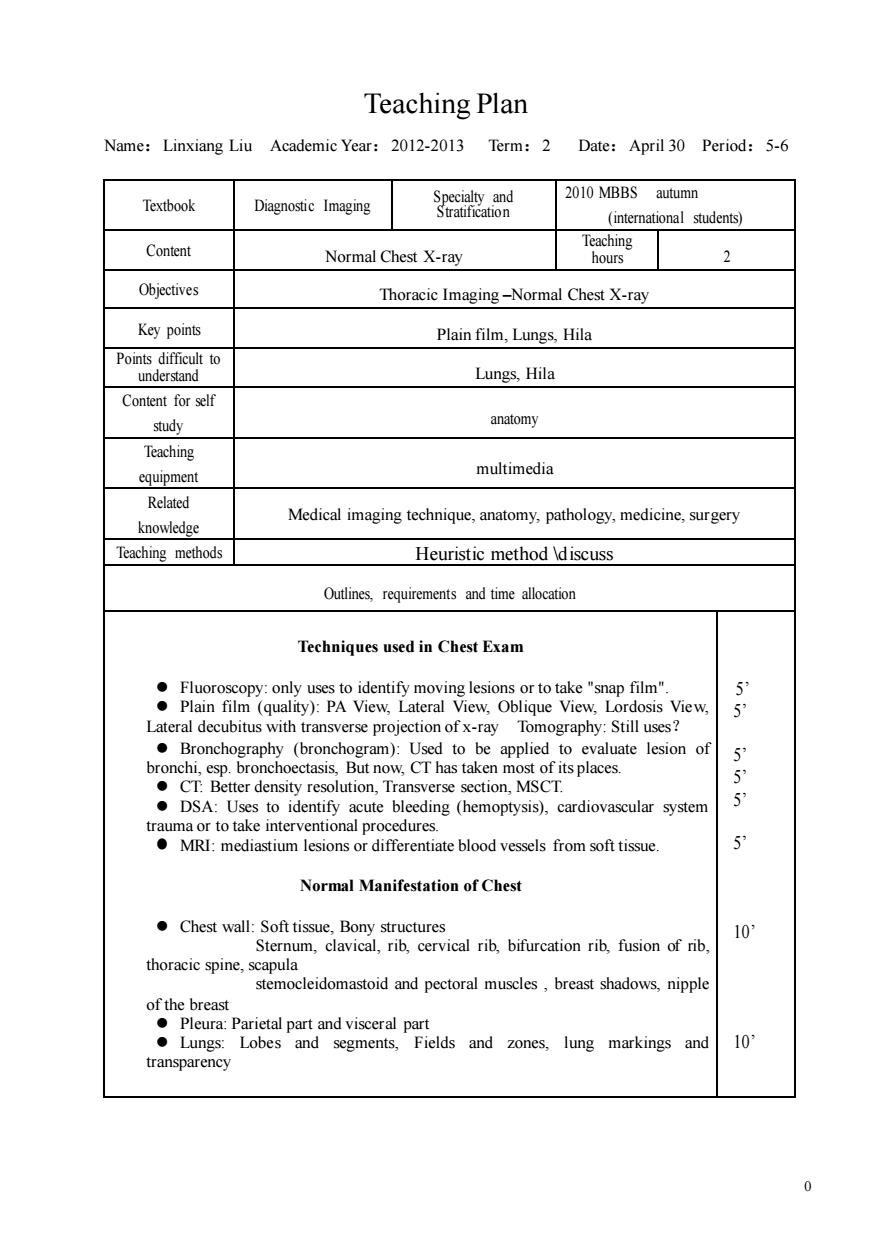
Teaching Plan Name:Linxiang Liu Academic Year:2012-2013 Term:2 Date:April 30 Period:5-6 Textbook Sautsaion 2010 MBBS autumn Diagnostic Imaging (international students) Content Normal Chest X-ray Teaching hours 2 Objectives Thoracic Imaging-Normal Chest X-ray Key points Plain film,Lungs,Hila Points difficult to understand Lungs,Hila Content for self study anatomy Teaching cquipment multimedia Related knowledge Medical imaging technique,anatomy,pathology,medicine,surgery Teaching methods Heuristic method \discuss Outlines requirements and time allocation Techniques used in Chest Exam is View, 5 ●Broncho m):Used to be valuate lesion of CT.Better density resolution,Transverse section,MSCT. DSA:Uses to identify acute bleeding (hemoptysis),cardiovascular system 5 rrRneeamisoasodireiaicblodvesesiomoAtiac Normal Manifestation of Chest Chest wall:Soft tissue.Bony structures 10 Sternum,clavical,rib,cervical rib,bifurcation rib,fusion of rib, thoracic spine,scapula stemocleidomastoid and pectoral muscles,breast shadows,nipple 色小地的tg segments 10 transparency 00 Teaching Plan Name:Linxiang Liu Academic Year:2012-2013 Term:2 Date:April 30 Period:5-6 Textbook Diagnostic Imaging Specialty and Stratification 2010 MBBS autumn (international students) Content Normal Chest X-ray Teaching hours 2 Objectives Thoracic Imaging –Normal Chest X-ray Key points Plain film, Lungs, Hila Points difficult to understand Lungs, Hila Content for self study anatomy Teaching equipment multimedia Related knowledge Medical imaging technique, anatomy, pathology, medicine, surgery Teaching methods Heuristic method \discuss Outlines, requirements and time allocation Techniques used in Chest Exam ⚫ Fluoroscopy: only uses to identify moving lesions or to take "snap film". ⚫ Plain film (quality): PA View, Lateral View, Oblique View, Lordosis View, Lateral decubitus with transverse projection of x-ray Tomography: Still uses? ⚫ Bronchography (bronchogram): Used to be applied to evaluate lesion of bronchi, esp. bronchoectasis, But now, CT has taken most of its places. ⚫ CT: Better density resolution, Transverse section, MSCT. ⚫ DSA: Uses to identify acute bleeding (hemoptysis), cardiovascular system trauma or to take interventional procedures. ⚫ MRI: mediastium lesions or differentiate blood vessels from soft tissue. Normal Manifestation of Chest ⚫ Chest wall: Soft tissue, Bony structures Sternum, clavical, rib, cervical rib, bifurcation rib, fusion of rib, thoracic spine, scapula stemocleidomastoid and pectoral muscles , breast shadows, nipple of the breast ⚫ Pleura: Parietal part and visceral part ⚫ Lungs: Lobes and segments, Fields and zones, lung markings and transparency 5’ 5’ 5’ 5’ 5’ 5’ 10’ 10’