正在加载图片...

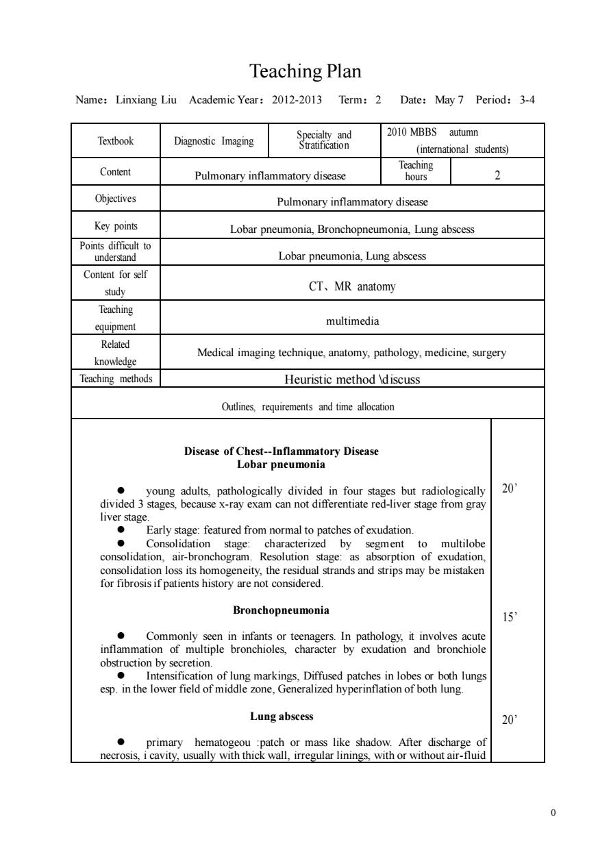
Teaching Plan Name:Linxiang Liu Academic Year:2012-2013 Term:2 Date:May7 Period:3-4 2010 MBBS autumn Textbook Diagnostic Imaging Suaion (students) Content Pulmonary inflammatory disease Teaching 2 Objectives Pulmonary inflammatory disease Key points Lobar pneumonia,Bronchopneumonia,Lung abscess Points difficult to Lobar pneumonia,Lung abscess Content for self study CT、MR anatomy Teaching equipment multimedia Related Medical imaging technique,anatomy,pathology,medicine,surgery knowledge Teaching methods Heuristic method discuss Outlines,requirements and time allocation atory Disease r pneumonia ● young adults,pathologically divided in four stages but radiologically 20 divided 3 stages,because x-ray exam can not differentiate red-liver stage from gray eoaimbRaenspon8e consolidation loss its homogeneity,the residual strands and strips may be mistaken for fibrosis if patients history are not considered. Bronchopneumonia 15 Commonly seen in infants or teenagers.In pathology it involves acute inflammation of multiple bronchioles,character by exudation and bronchiole obstruction by secretion. in lob Lung abscess 20 primary hematogeou :patch or mass like shadow.After discharge of necrosis,i cavity.usually with thick wall,irregular linings,with or without air-fluid 0 0 Teaching Plan Name:Linxiang Liu Academic Year:2012-2013 Term:2 Date:May 7 Period:3-4 Textbook Diagnostic Imaging Specialty and Stratification 2010 MBBS autumn (international students) Content Pulmonary inflammatory disease Teaching hours 2 Objectives Pulmonary inflammatory disease Key points Lobar pneumonia, Bronchopneumonia, Lung abscess Points difficult to understand Lobar pneumonia, Lung abscess Content for self study CT、MR anatomy Teaching equipment multimedia Related knowledge Medical imaging technique, anatomy, pathology, medicine, surgery Teaching methods Heuristic method \discuss Outlines, requirements and time allocation Disease of Chest-Inflammatory Disease Lobar pneumonia ⚫ young adults, pathologically divided in four stages but radiologically divided 3 stages, because x-ray exam can not differentiate red-liver stage from gray liver stage. ⚫ Early stage: featured from normal to patches of exudation. ⚫ Consolidation stage: characterized by segment to multilobe consolidation, air-bronchogram. Resolution stage: as absorption of exudation, consolidation loss its homogeneity, the residual strands and strips may be mistaken for fibrosis if patients history are not considered. Bronchopneumonia ⚫ Commonly seen in infants or teenagers. In pathology, it involves acute inflammation of multiple bronchioles, character by exudation and bronchiole obstruction by secretion. ⚫ Intensification of lung markings, Diffused patches in lobes or both lungs esp. in the lower field of middle zone, Generalized hyperinflation of both lung. Lung abscess ⚫ primary hematogeou :patch or mass like shadow. After discharge of necrosis, i cavity, usually with thick wall, irregular linings, with or without air-fluid 20’ 15’ 20’