正在加载图片...

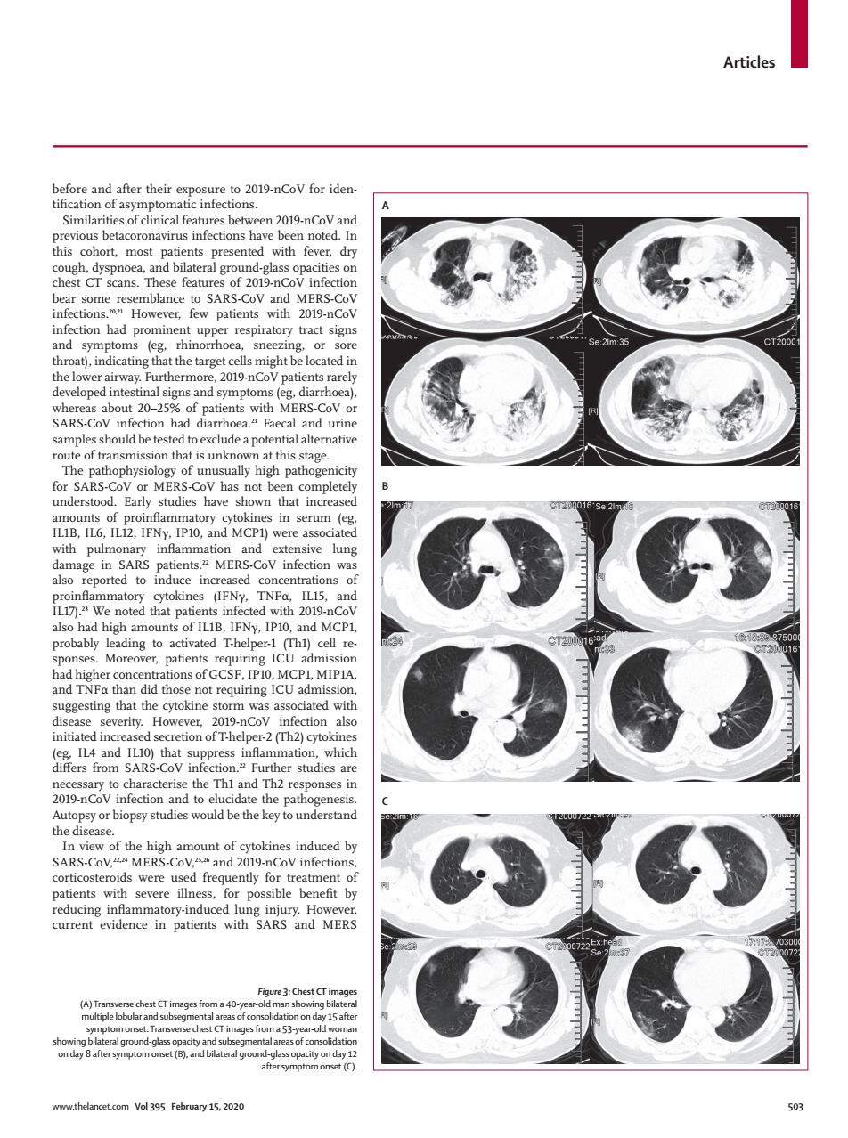
Articles hsomondanpeR20i9-ncovforiden Smilaitsofdalfeanscsbete209nCaN1nd nd bil t si hoe rairway.Furthermore.2019-nCoV patients ra signs and symptoms SARS-CoV ut20- Faec MEp of unus igh patho Early studies shown that increase wi pulm lso nd F,IP1( stine that the cytokine storm was ass iated wit disease severity. 10 whicl haracterise the ced b corticosteroids were u quently for treatme Figuve Chest CT image a40- Vol395 February 15.202Articles www.thelancet.com Vol 395 February 15, 2020 503 before and after their exposure to 2019-nCoV for iden - tification of asymptomatic infections. Similarities of clinical features between 2019-nCoV and previous betacoronavirus infections have been noted. In this cohort, most patients presented with fever, dry cough, dyspnoea, and bilateral ground-glass opacities on chest CT scans. These features of 2019-nCoV infection bear some resemblance to SARS-CoV and MERS-CoV infections.20,21 However, few patients with 2019-nCoV infection had prominent upper respiratory tract signs and symptoms (eg, rhinorrhoea, sneezing, or sore throat), indicating that the target cells might be located in the lower airway. Furthermore, 2019-nCoV patients rarely developed intestinal signs and symptoms (eg, diarrhoea), whereas about 20–25% of patients with MERS-CoV or SARS-CoV infection had diarrhoea.21 Faecal and urine samples should be tested to exclude a potential alternative route of transmission that is unknown at this stage. The pathophysiology of unusually high pathogenicity for SARS-CoV or MERS-CoV has not been completely understood. Early studies have shown that increased amounts of proinflammatory cytokines in serum (eg, IL1B, IL6, IL12, IFNγ, IP10, and MCP1) were associated with pulmonary inflammation and extensive lung damage in SARS patients.22 MERS-CoV infection was also reported to induce increased concentrations of proinflammatory cytokines (IFNγ, TNFα, IL15, and IL17).23 We noted that patients infected with 2019-nCoV also had high amounts of IL1B, IFNγ, IP10, and MCP1, probably leading to activated T-helper-1 (Th1) cell re - sponses. Moreover, patients requiring ICU admission had higher concentrations of GCSF, IP10, MCP1, MIP1A, and TNFα than did those not requiring ICU admission, suggesting that the cytokine storm was associated with disease severity. However, 2019-nCoV infection also initiated increased secretion of T-helper-2 (Th2) cytokines (eg, IL4 and IL10) that suppress inflammation, which differs from SARS-CoV infection.22 Further studies are necessary to characterise the Th1 and Th2 responses in 2019-nCoV infection and to elucidate the pathogenesis. Autopsy or biopsy studies would be the key to understand the disease. In view of the high amount of cytokines induced by SARS-CoV, 22,24 MERS-CoV,25,26 and 2019-nCoV infections, corticosteroids were used frequently for treatment of patients with severe illness, for possible benefit by reducing inflammatory-induced lung injury. However, current evidence in patients with SARS and MERS Figure 3: Chest CT images (A) Transverse chest CT images from a 40-year-old man showing bilateral multiple lobular and subsegmental areas of consolidation on day 15 after symptom onset. Transverse chest CT images from a 53-year-old woman showing bilateral ground-glass opacity and subsegmental areas of consolidation on day 8 after symptom onset (B), and bilateral ground-glass opacity on day 12 after symptom onset (C). ABC