正在加载图片...
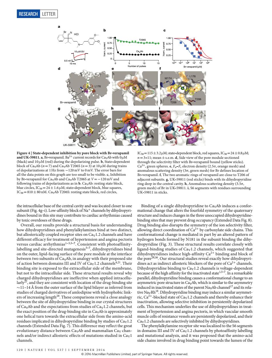
RESEARCH LETTER b re 4 Sta e-dep dent inhibition by pore block with B =115±32 state- ,1C0=24±0.8M vith Br -120mV g de sity (30 h)for all to be visible se to T206o ions s in b.CavAb: ()bnd dihydoon boun etry of the o mediated in pa t by an alte with their the pore ural st es rev exactly ho 产 s is volta g-dependen a rem Ab studies of chargedde similar asvm nism un ells of res sta ly depolari d.h (eTe ine r and mutatic sis and it was r bd that the amino- channels. side bndin pont towards themen of the 120 I NATURE I VOL 537 II 2016 Nature.All rights reserve RESEARCH Letter 120 | NA T U RE | VOL 537 | 1 september 2016 the intracellular base of the central cavity and was located closer to one subunit (Fig. 4g–i). Low-affinity block of Na+ channels by dihydropyridines bound in this site may contribute to cardiac arrhythmias caused by toxic overdoses of these drugs. Overall, our results provide a structural basis for understanding how dihydropyridines and phenylalkylamines bind at two distinct, but allosterically coupled receptor sites on CaV1.2 channels and have different efficacy for treatment of hypertension and angina pectoris versus cardiac arrhythmias1–3,5–7. Consistent with photoaffinitylabelling and site-directed mutagenesis5–7, dihydropyridines bind on the outer, lipid-facing surface of the pore module at the interface between two subunits of CaVAb, in analogy with their proposed site of action between domains III and IV of CaV1.2 channels4,21. Their binding site is exposed to the extracellular side of the membrane, but not to the intracellular side. These structural results reveal why charged dihydropyridines are ineffective when applied intracellularly27, and they are consistent with location of the drug-binding site ~11–14Å from the outer surface of the lipid bilayer as inferred from studies of charged derivatives of amlodipine with hydrophobic linkers of increasing length28. These comparisons reveal a close analogy between the site of dihydropyridine binding in our crystal structures of CaVAb and the expectations from studies of CaV1.2 channels, but the exact position of the drug-binding site in CaVAb is approximately one helical turn towards the extracellular side from the amino-acid residues implicated in dihydropyridine binding by studies of CaV1.2 channels (Extended Data Fig. 7). This difference may reflect the great evolutionary distance between CaVAb and mammalian CaV channels and/or indirect allosteric effects of mutations studied in CaV1 channels. Binding of a single dihydropyridine to CaVAb induces a conformational change that alters the fourfold symmetry of the quaternary structure and induces changes in the three unoccupied dihydropyridinebinding sites that may prevent drug occupancy (Extended Data Fig. 8). Drug binding also disrupts the symmetry of the ion selectivity filter, allowing direct coordination of Ca2+ by carboxylate side chains. This conformational change is mediated in part by an altered pattern of hydrogen bonds formed by N181 in the subunit binding the dihydropyridine (Fig. 3). These structural results correlate closely with ligand-binding studies of CaV1.2 channels, which suggested that dihydropyridines induce high-affinity Ca2+ binding and block of the pore29,30. Our structural studies reveal exactly how dihydropyridines act as indirect allosteric blockers of the pore of Ca2+ channels. Dihydropyridine binding to CaV1.2 channels is voltage-dependent because of the high affinity for the inactivated state1,5–7. In a remarkable parallel, dihydropyridine binding causes a conformational change to an asymmetric pore structure in CaVAb, which is similar to the asymmetry induced in inactivated states of the parent NaVAb channel17 and its relative NaVRh18. Dihydropyridine binding may induce a similar asymmetric, Ca2+-blocked state of CaV1.2 channels and thereby enhance their inactivation, allowing selective inhibition in persistently depolarized cells. This mechanism underlies the use of dihydropyridines in treatment of hypertension and angina pectoris, in which vascular smooth muscle cells of resistance vessels are persistently depolarized, and their CaV1.2 channels are selectively inhibited by dihydropyridines. The phenylalkylamine receptor site was localized to the S6 segments in domains III and IV of CaV1.2 channels by photoaffinity labelling and mutational analysis, and it was proposed that the amino-acid side chains involved in drug binding point towards the lumen of the 10 ms 0.5 nA 10 μM BV b c T206S 10–2 10–1 100 101 102 103 Br-verapamil (μM) a ° ° S6 S6 V210 I213 M209 I213 T206 V210 M209 I213 V210 V210 I213 UK-59811 L176 T206 M174 L176 T206 Br-verapamil T206 d e f g h i CaVAb O O N Br N HCl 0.25 0.50 0.75 1.00 0.25 0.50 0.75 1.00 0 Norm. current Norm. current 0 5 10 15 Number of pulses Figure 4 | State-dependent inhibition by pore block with Br-verapamil and UK-59811. a, Br-verapamil. Ba2+ current records for CaVAb with 0μM (black) and 10 μM (red) during the depolarizing pulse. b, State-dependent block of CaVAb (n=7) and CaVAb T206S (n=3) at 10 μM during trains of depolarizations at 1Hz from −120 mV to 0 mV. The error bars for all the data points on this graph are too small to be visible. c, Inhibition by Br-verapamil for CaVAb and CaVAb T206S at V=−120 mV and following trains of depolarizations as in b. CaVAb: resting state block, blue circles, IC50=24±1.6 μM; state-dependent block, blue squares, IC50=810±80 nM. CaVAb T206S: resting state block, red circles, IC50=115±3.2μM; state-dependent block, red squares, IC50=24±0.8μM; n=3–11; mean±s.e.m. d, Side view of the pore module sectioned through the selectivity filter with Br-verapamil bound (yellow sticks). Ca2+, green spheres. e, Fo–Fc electron density (2.5σ, orange mesh) and anomalous scattering density (3σ, green mesh) for Br defines location of Br-verapamil. f, The two aromatic rings of verapamil are close to T206 of adjacent subunits. g, UK-59811 (red sticks) binds with its dihydropyridine ring deep in the central cavity. h, Anomalous scattering density (3.5σ, green mesh) of Br in UK-59811. i, S6 segments with residues surrounding UK-59811 in sticks. © 2016 Macmillan Publishers Limited, part of Springer Nature. All rights reserved