正在加载图片...
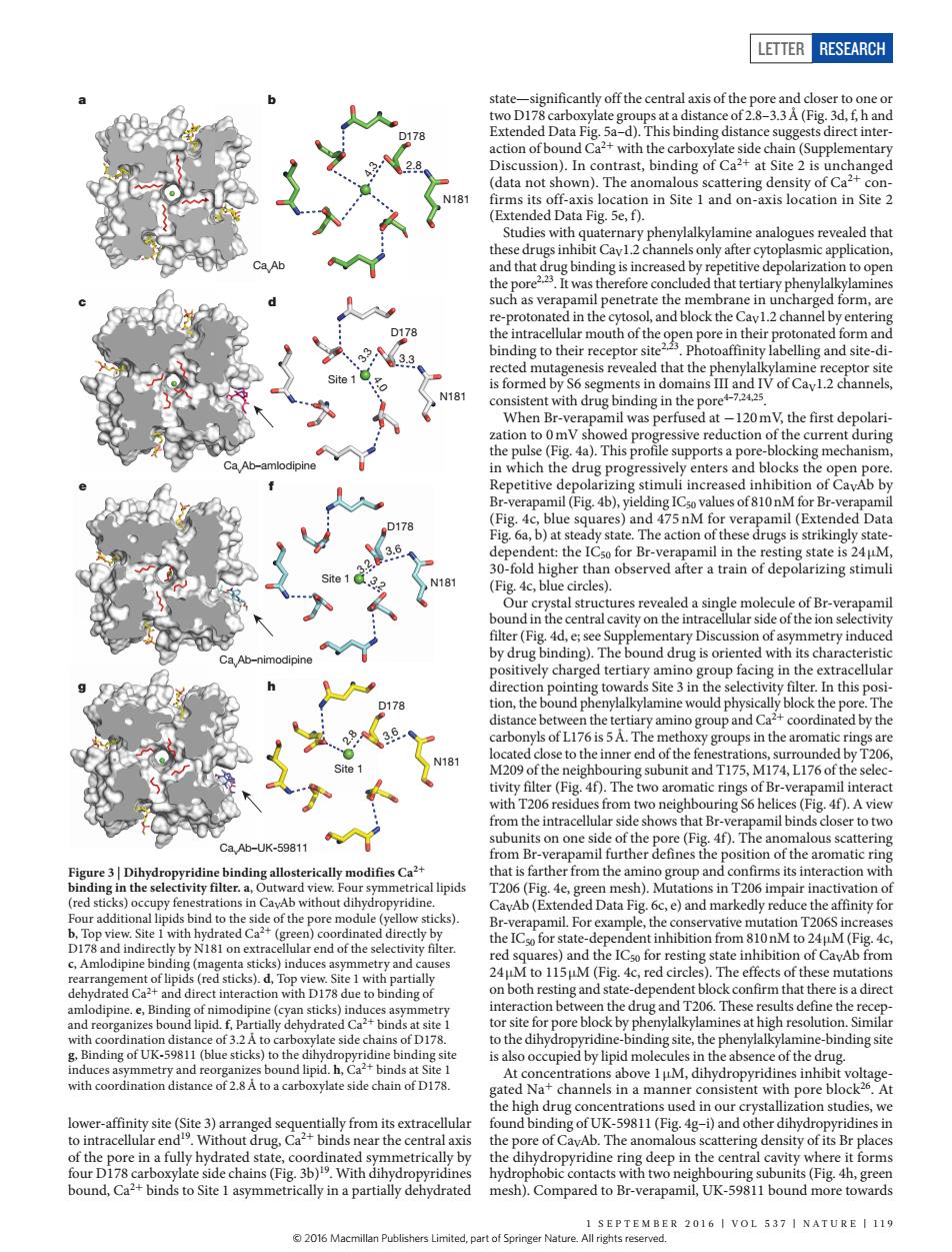
LETTER RESEARCH icantly off the c atter 0naioataase1andom-adslocaioninsie2 and block the 2 nV,the first de ssive reduction o which the d ckh四 Detitive 1 charged tertiary ar 5 i up facing in the e coordinate Iby th of Br-ve ifics ca? nof CavAb from te l wi e4 ing and state fUK-5981 (stickay n the phenylakylmine-ndinge e high drug con used in ourc ing of U mesh Compared to Br-verapUK-s98 pound SEPTEMBER 2016 I VOL 537 I NATURE I119Letter RESEARCH 1 september 2016 | VOL 537 | NA T U RE | 119 lower-affinity site (Site 3) arranged sequentially from its extracellular to intracellular end19. Without drug, Ca2+ binds near the central axis of the pore in a fully hydrated state, coordinated symmetrically by four D178 carboxylate side chains (Fig. 3b) 19. With dihydropyridines bound, Ca2+ binds to Site 1 asymmetrically in a partially dehydrated state—significantly off the central axis of the pore and closer to one or two D178 carboxylate groups at a distance of 2.8–3.3Å (Fig. 3d, f, h and Extended Data Fig. 5a–d). This binding distance suggests direct interaction of bound Ca2+ with the carboxylate side chain (Supplementary Discussion). In contrast, binding of Ca2+ at Site 2 is unchanged (data not shown). The anomalous scattering density of Ca2+ confirms its off-axis location in Site 1 and on-axis location in Site 2 (Extended Data Fig. 5e, f). Studies with quaternary phenylalkylamine analogues revealed that these drugs inhibit CaV1.2 channels only after cytoplasmic application, and that drug binding is increased by repetitive depolarization to open the pore2,23. It was therefore concluded that tertiary phenylalkylamines such as verapamil penetrate the membrane in uncharged form, are re-protonated in the cytosol, and block the CaV1.2 channel by entering the intracellular mouth of the open pore in their protonated form and binding to their receptor site2,23. Photoaffinity labelling and site-directed mutagenesis revealed that the phenylalkylamine receptor site is formed by S6 segments in domains III and IV of CaV1.2 channels, consistent with drug binding in the pore4–7,24,25. When Br-verapamil was perfused at −120mV, the first depolarization to 0mV showed progressive reduction of the current during the pulse (Fig. 4a). This profile supports a pore-blocking mechanism, in which the drug progressively enters and blocks the open pore. Repetitive depolarizing stimuli increased inhibition of CaVAb by Br-verapamil (Fig. 4b), yielding IC50 values of 810nM for Br-verapamil (Fig. 4c, blue squares) and 475 nM for verapamil (Extended Data Fig. 6a, b) at steady state. The action of these drugs is strikingly statedependent: the IC50 for Br-verapamil in the resting state is 24 μM, 30-fold higher than observed after a train of depolarizing stimuli (Fig. 4c, blue circles). Our crystal structures revealed a single molecule of Br-verapamil bound in the central cavity on the intracellular side of the ion selectivity filter (Fig. 4d, e; see Supplementary Discussion of asymmetry induced by drug binding). The bound drug is oriented with its characteristic positively charged tertiary amino group facing in the extracellular direction pointing towards Site 3 in the selectivity filter. In this position, the bound phenylalkylamine would physically block the pore. The distance between the tertiary amino group and Ca2+ coordinated by the carbonyls of L176 is 5Å. The methoxy groups in the aromatic rings are located close to the inner end of the fenestrations, surrounded by T206, M209 of the neighbouring subunit and T175, M174, L176 of the selectivity filter (Fig. 4f). The two aromatic rings of Br-verapamil interact with T206 residues from two neighbouring S6 helices (Fig. 4f). A view from the intracellular side shows that Br-verapamil binds closer to two subunits on one side of the pore (Fig. 4f). The anomalous scattering from Br-verapamil further defines the position of the aromatic ring that is farther from the amino group and confirms its interaction with T206 (Fig. 4e, green mesh). Mutations in T206 impair inactivation of CaVAb (Extended Data Fig. 6c, e) and markedly reduce the affinity for Br-verapamil. For example, the conservative mutation T206S increases the IC50 for state-dependent inhibition from 810nM to 24μM (Fig. 4c, red squares) and the IC50 for resting state inhibition of CaVAb from 24μM to 115μM (Fig. 4c, red circles). The effects of these mutations on both resting and state-dependent block confirm that there is a direct interaction between the drug and T206. These results define the receptor site for pore block by phenylalkylamines at high resolution. Similar to the dihydropyridine-binding site, the phenylalkylamine-binding site is also occupied by lipid molecules in the absence of the drug. At concentrations above 1μM, dihydropyridines inhibit voltagegated Na+ channels in a manner consistent with pore block26. At the high drug concentrations used in our crystallization studies, we found binding of UK-59811 (Fig. 4g–i) and other dihydropyridines in the pore of CaVAb. The anomalous scattering density of its Br places the dihydropyridine ring deep in the central cavity where it forms hydrophobic contacts with two neighbouring subunits (Fig. 4h, green mesh). Compared to Br-verapamil, UK-59811 bound more towards 2.8 3.6 D178 N181 Site 1 f D178 N181 3.2 3.6 3.2 Site 1 3.3 4.0 D178 N181 Site 1 d b CavAb–amlodipine CavAb CavAb–nimodipine CavAb–UK-59811 a c e g h 4.3 2.8 D178 N181 3.3 Figure 3 | Dihydropyridine binding allosterically modifies Ca2+ binding in the selectivity filter. a, Outward view. Four symmetrical lipids (red sticks) occupy fenestrations in CaVAb without dihydropyridine. Four additional lipids bind to the side of the pore module (yellow sticks). b, Top view. Site 1 with hydrated Ca2+ (green) coordinated directly by D178 and indirectly by N181 on extracellular end of the selectivity filter. c, Amlodipine binding (magenta sticks) induces asymmetry and causes rearrangement of lipids (red sticks). d, Top view. Site 1 with partially dehydrated Ca2+ and direct interaction with D178 due to binding of amlodipine. e, Binding of nimodipine (cyan sticks) induces asymmetry and reorganizes bound lipid. f, Partially dehydrated Ca2+ binds at site 1 with coordination distance of 3.2Å to carboxylate side chains of D178. g, Binding of UK-59811 (blue sticks) to the dihydropyridine binding site induces asymmetry and reorganizes bound lipid. h, Ca2+ binds at Site 1 with coordination distance of 2.8Å to a carboxylate side chain of D178. © 2016 Macmillan Publishers Limited, part of Springer Nature. All rights reserved