正在加载图片...

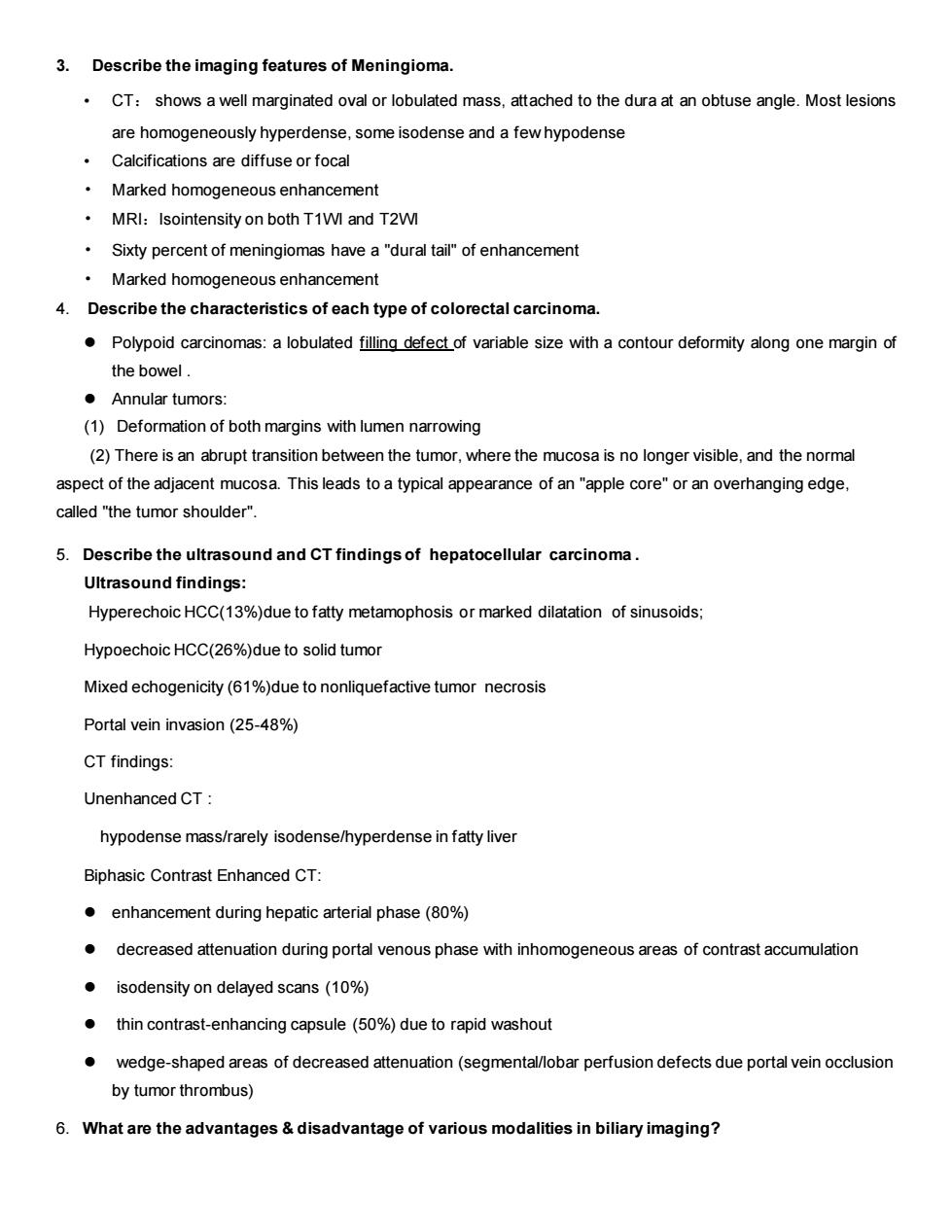
3.Describe the imaging features of Meningioma CT:shows a well marginated oval or lobulated mass,attached to the dura at an obtuse angle.Most lesions are homogeneously hyperdense.some isodense and a few hypodense Calcifications are diffuse or focal Marked homogeneous enhancement MRI:Isointensity on both T1WI and T2WI Sixty percent of meningiomas have a"dural tail"of enhancement Marked homogeneous enhancement 4.Describe the characteristics of each type of colorectal carcinoma. Polypoid carcinomas:a lobulated filling defect of variable size with a contour deformity along one margin of the bowel Annular tumors: (1)Deformation of both margins with lumen narrowing (2)There is an abrupt transition between the tumor,where the mucosa is no longer visible,and the normal aspect of the adjacent mucosa.This leads to a typical appearance of an"apple core"or an overhanging edge. called "the tumor shoulder". 5.Describe the ultrasound and CT findings of hepatocellular carcinoma. Ultrasound findings Hyperechoic HCC(13%)due to fatty metamophosis or marked dilatation of sinusoids Hypoechoic HCC(26%)due to solid tumor Mixed echogenicity(61%)due to nonliquefactive tumor necrosis Portal vein invasion(25-48%) CT findings: Unenhanced CT: hypodense mass/rarely isodense/hyperdense in fatty liver Biphasic Contrast Enhanced CT: enhancement during hepatic arterial phase(80%) decreased attenuation during portal venous phase with inhomogeneous areas of contrast accumulation isodensity on delayed scans(10%) thin contrast-enhancing capsule(50%)due to rapid washout wedge-shaped areas of decreased attenuation(segmental/lobar perfusion defects due portal vein occlusion by tumor thrombus) 6.What are the advantages disadvantage of various modalities in biliary imaging?3. Describe the imaging features of Meningioma. • CT: shows a well marginated oval or lobulated mass, attached to the dura at an obtuse angle. Most lesions are homogeneously hyperdense, some isodense and a few hypodense • Calcifications are diffuse or focal • Marked homogeneous enhancement • MRI:Isointensity on both T1WI and T2WI • Sixty percent of meningiomas have a "dural tail" of enhancement • Marked homogeneous enhancement 4. Describe the characteristics of each type of colorectal carcinoma. ⚫ Polypoid carcinomas: a lobulated filling defect of variable size with a contour deformity along one margin of the bowel . ⚫ Annular tumors: (1) Deformation of both margins with lumen narrowing (2) There is an abrupt transition between the tumor, where the mucosa is no longer visible, and the normal aspect of the adjacent mucosa. This leads to a typical appearance of an "apple core" or an overhanging edge, called "the tumor shoulder". 5. Describe the ultrasound and CT findings of hepatocellular carcinoma . Ultrasound findings: Hyperechoic HCC(13%)due to fatty metamophosis or marked dilatation of sinusoids; Hypoechoic HCC(26%)due to solid tumor Mixed echogenicity (61%)due to nonliquefactive tumor necrosis Portal vein invasion (25-48%) CT findings: Unenhanced CT : hypodense mass/rarely isodense/hyperdense in fatty liver Biphasic Contrast Enhanced CT: ⚫ enhancement during hepatic arterial phase (80%) ⚫ decreased attenuation during portal venous phase with inhomogeneous areas of contrast accumulation ⚫ isodensity on delayed scans (10%) ⚫ thin contrast-enhancing capsule (50%) due to rapid washout ⚫ wedge-shaped areas of decreased attenuation (segmental/lobar perfusion defects due portal vein occlusion by tumor thrombus) 6. What are the advantages & disadvantage of various modalities in biliary imaging?