正在加载图片...
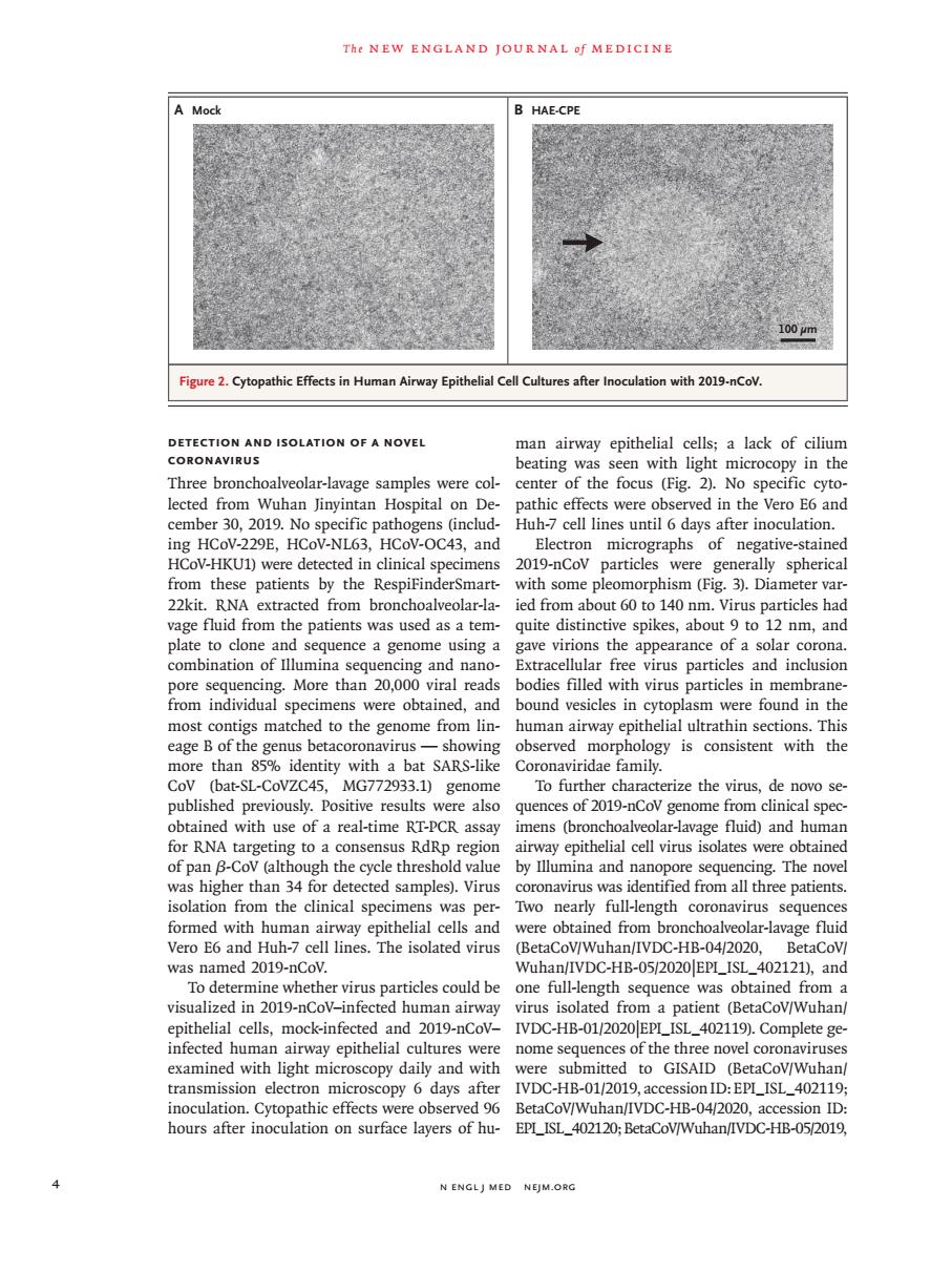
Thr NEW ENGLAND JOURNAL Of MEDICINE A Mock 100m 2.0 athic Efferts in H ocuation with 19-nCoV DETECTION AND ISOLATION OF A NOVEL man airway epithelial cells:a lack of ciliun CORONAVIRUS beating was seen with light microcopy in the Three bronchoalveolar-lavage samples were col- center of the focus (Fig.2).No specific cyto- lected from Wuhan Jinyintan Hospital on De pathic effects were observed in the Vero E and cember 30,2019. Huh-7 cell lines until 6 days after inoculation 43,and 2019c negative-s 。 patients by the Resp with some phism 22kit RNA extracted from bronchoalyeolar-la ied from about 60 to 140 nm.Virus par ticles ha rage fluid from the patients was used as a tem quite distinctive spikes,about 9 to 12 nm,and plate to clone and sequence a genome using a gave virions the appearance of a solar corona combination of lllumina Extracellular free virus particles and inclusio d sicles in particles in me ed to the e found nhology is tent more than 85%identity with a bat SARS-like Coronaviridae family CoV (bat-SL-CoVZC45,MG772933.1)genome To further characterize the virus,de novo se- published previously.Positive results ere alsc quences of 2019-nCov genome from clinical spec imens (bronchoalv age fluid)and humar RN e tes we of pan p isolation from the clinical specimens was per Tvo nearly full-length coronavirus sequence formed with human airwa epithelial cells and were obtained from hronchoalyeolar-lavage fluid Vero E6 and Huh-7 cell lines.The isolated virus (BetaCoV/Wuhan/IVDC-HB-04/2020, BetaCoV was named 2019-n Wuhan/IVDC-HB-05/2020EPI_ISL_402 21),and insparticdeseouldbs one ful engt sequenc obtained rom mock-infa IVDC-HB-0120 the th examined with light microscopy daily and with were submitted to GISAID (BetaCov/Wuhan transmission electron microscopy 6days after IVDC-HB-01/2019,accessionID:EPI_ISL_402119 noculation.Cytopathic effects were observec BetaCoV/Wuhan/IVDC-HB-04/2020,accession ID nours after inoculation on surface layers of hu EPI_ISL_402120;BetaCoV/Wuhan/IVDC-HB-05/2019 N ENGLJ MED NEJM.ORG4 n engl j med nejm.org The new england journal o f medicine Detection and Isolation of a Novel Coronavirus Three bronchoalveolar-lavage samples were collected from Wuhan Jinyintan Hospital on December 30, 2019. No specific pathogens (including HCoV-229E, HCoV-NL63, HCoV-OC43, and HCoV-HKU1) were detected in clinical specimens from these patients by the RespiFinderSmart- 22kit. RNA extracted from bronchoalveolar-lavage fluid from the patients was used as a template to clone and sequence a genome using a combination of Illumina sequencing and nanopore sequencing. More than 20,000 viral reads from individual specimens were obtained, and most contigs matched to the genome from lineage B of the genus betacoronavirus — showing more than 85% identity with a bat SARS-like CoV (bat-SL-CoVZC45, MG772933.1) genome published previously. Positive results were also obtained with use of a real-time RT-PCR assay for RNA targeting to a consensus RdRp region of pan β-CoV (although the cycle threshold value was higher than 34 for detected samples). Virus isolation from the clinical specimens was performed with human airway epithelial cells and Vero E6 and Huh-7 cell lines. The isolated virus was named 2019-nCoV. To determine whether virus particles could be visualized in 2019-nCoV–infected human airway epithelial cells, mock-infected and 2019-nCoV– infected human airway epithelial cultures were examined with light microscopy daily and with transmission electron microscopy 6 days after inoculation. Cytopathic effects were observed 96 hours after inoculation on surface layers of human airway epithelial cells; a lack of cilium beating was seen with light microcopy in the center of the focus (Fig. 2). No specific cytopathic effects were observed in the Vero E6 and Huh-7 cell lines until 6 days after inoculation. Electron micrographs of negative-stained 2019-nCoV particles were generally spherical with some pleomorphism (Fig. 3). Diameter varied from about 60 to 140 nm. Virus particles had quite distinctive spikes, about 9 to 12 nm, and gave virions the appearance of a solar corona. Extracellular free virus particles and inclusion bodies filled with virus particles in membranebound vesicles in cytoplasm were found in the human airway epithelial ultrathin sections. This observed morphology is consistent with the Coronaviridae family. To further characterize the virus, de novo sequences of 2019-nCoV genome from clinical specimens (bronchoalveolar-lavage fluid) and human airway epithelial cell virus isolates were obtained by Illumina and nanopore sequencing. The novel coronavirus was identified from all three patients. Two nearly full-length coronavirus sequences were obtained from bronchoalveolar-lavage fluid (BetaCoV/Wuhan/IVDC-HB-04/2020, BetaCoV/ Wuhan/IVDC-HB-05/2020|EPI_ISL_402121), and one full-length sequence was obtained from a virus isolated from a patient (BetaCoV/Wuhan/ IVDC-HB-01/2020|EPI_ISL_402119). Complete genome sequences of the three novel coronaviruses were submitted to GISAID (BetaCoV/Wuhan/ IVDC-HB-01/2019, accession ID: EPI_ISL_402119; BetaCoV/Wuhan/IVDC-HB-04/2020, accession ID: EPI_ISL_402120; BetaCoV/Wuhan/IVDC-HB-05/2019, Figure 2. Cytopathic Effects in Human Airway Epithelial Cell Cultures after Inoculation with 2019-nCoV. A Mock B HAE-CPE 100 µm