正在加载图片...
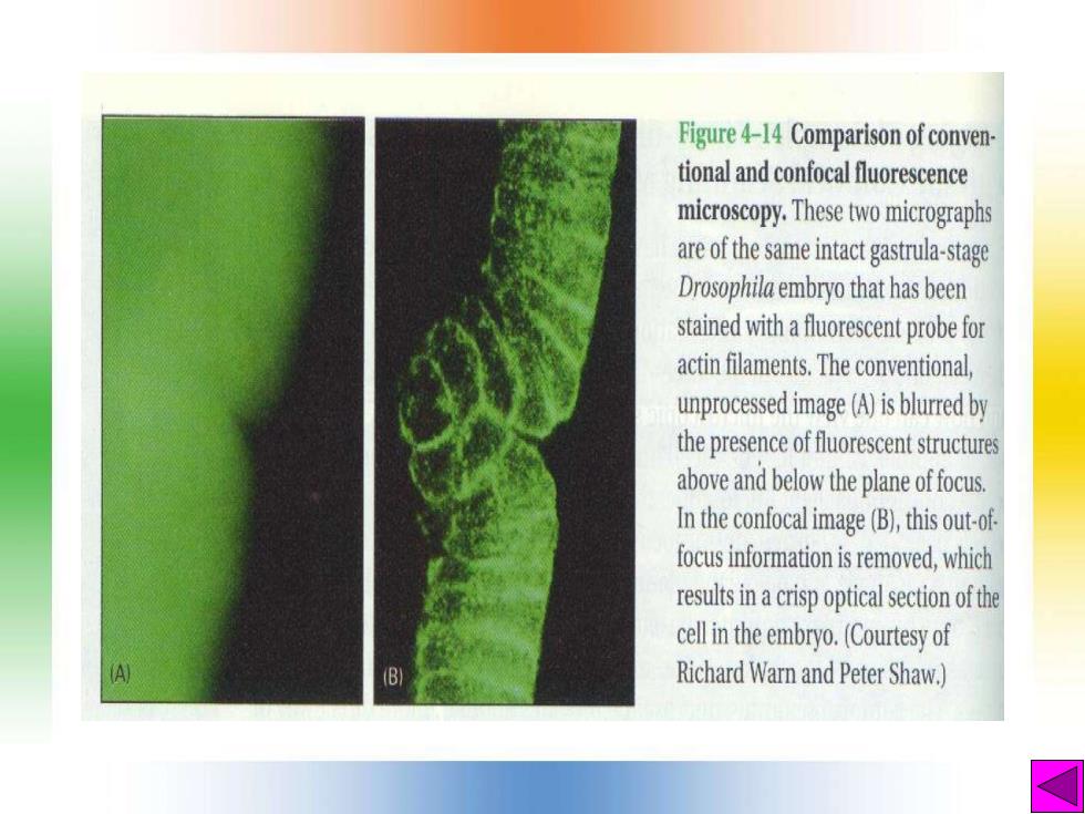
Figure 4-14 Comparison of conven- tional and confocal fluorescence microscopy.These two micrographs are of the same intact gastrula-stage Drosophila embryo that has been stained with a fuorescent probe for actin filaments.The conventional, unprocessed image (A)is blurred by the presence of fuoresetsructures above and below the plane of focus. In the confocaimage (B),this out-of focus information is removed,which resuiacrispopticlscofthe cell in the embryo.(Courtesy of (B) Richard Warn and Peter Shaw.)