正在加载图片...
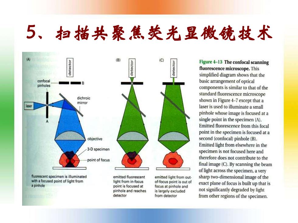
5、扫描共聚焦荧光显微镜技术 (B) Figure 4-13 The confocal scanning fluorescence microscope.This simplified diagram shows that the confocal basic arrangement of optical pinholes components is similar to that of the standard fluorescence microscope dichroic mirror shown in Figure 4-7 except that a laser is used to illuminate a small pinhole whose image is focused at a single point in the specimen (A). Emitted fluorescence from this focal point in the specimen is focused at a objective second (confocal)pinhole (B). Emitted light from elsewhere in the 3-D specimen specimen is not focused here and point of focus therefore does not contribute to the final image(C).By scanning the beam of light across the specimen,a very fluorescent specimen is illuminated emitted fluorescent emitted light from out. sharp two-dimensional image of the with a focused point of light from light from in-focus of-focus point is out of a pinhole point is focused at exact plane of focus is built up that is focus at pinhole and pinhole and reaches is largely excluded not significantly degraded by light detector from detector from other regions of the specimen.5、扫描共聚焦荧光显微镜技术