正在加载图片...

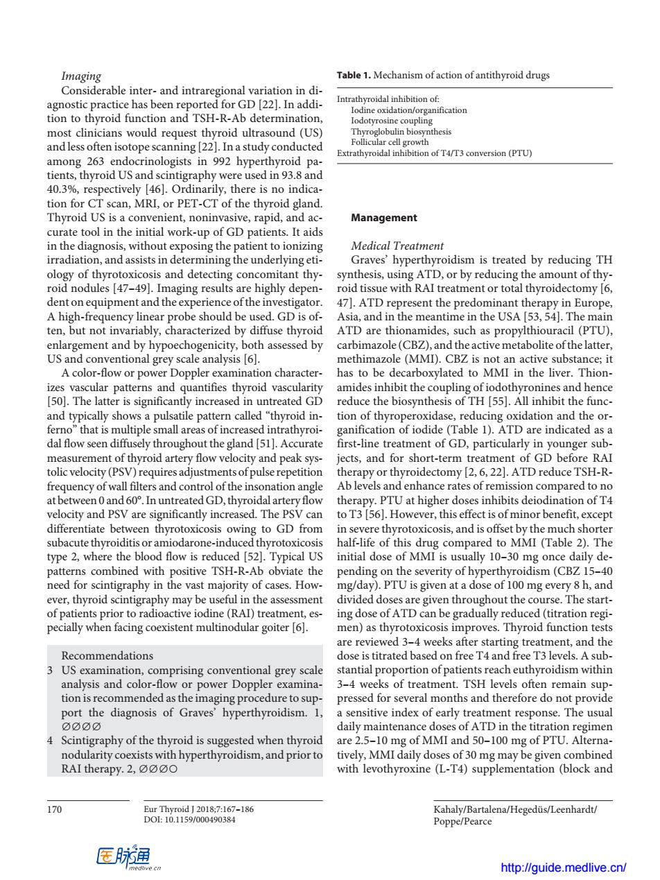
Imaging Table 1.Mechanism of action of antithyroid drugs Considerable inter-and intraregional variation in di- for GD [22].In addi- Intrathyroidal inhibition of: ne ox tio m0 reque soun ten is (Us) ay con 9p92hc93.8 ofT4/T3 conversion (PTU) oid US 40.3%,respectively 6.Ordinarily,there is no indica tion for CT scan,MRI,or PET-CT of the thyroid gland. Thyroid US is a convenient,noninvasive,rapid,and ac- Management curate tool in the initial work-up of GD patients.It aids Medical Treatment rradiat ying e sm is treated by reducing TH and det cting con thy D,or by red acing the ar nt of thy dthe ATD A high-freau used.GD is of Asia and in the m nti me in the USA 53.541.The ma ATD are thionamides,such as propylthiouracil (PTU). enlargement and by hypoechogenicity,both assessed by carbimazole(CBZ),and the active metabolite ofthe latter. US and conventional grey scale analysis [6] methimazole (MMI).CBZ is not an active substance;it A color-flow or po er Dopple examination character nas to be decarboxylated to MMI in the liver. it the coupling o is sign rea the biosyn :l hy 11 11 dal flow seen diffusely throughout the gland [51.Accurate first-line treatment of GD, OU measurement of thyroid artery flow velocity and peak sys tolic velocity(PSV)requires adjustments ofpulser repetition the frequency of wall filters and control of the insonation angle at.In untreatedGD,thyroidal artery flow therapy.PI U at higher doses inhibits deiodination of T are significantly increa can to T3 [56 How this effect is en thyr and is 2)Te 01 type 30 ombined with yate the the s pidism (CBZ 15 ever,thyroid scintig aphy may be useful in the assessment divided doses are given throughout the course.The start- of patients prior to radioactive iodine(RADtreatment,es- ing dose ofATD can be gradually reduced (titration regi- pecially when facing coexistent multinodular goiter [6]. men)as thyrotoxicosis improves.Thyroid function tes re reviewed. 4 weeks atter star ommendations titrated ati comprising conv al grey scale ntia op nded asthowe opp edu months nd the do e nsitive index of early treatment re e.The usual daily maintenance doses of ATD in the titration 4 Scintigraphy of the thyroid is suggested when thyroid are 2.5-10 mg of MMI and 50-100 mg of PTU.Alterna- nodularity coexists with hyperthyroidism,and prior to tively,MMI daily doses of 30 mg may be given combinec RAI therapy.2,oo with levothyroxine (L-T4)supplementation (block and Kahaly/Bartalena/Hegeduis/Leenhardt/ Poppe/Pearce 医通 http://guide.medlive.cn/ Kahaly/Bartalena/Hegedüs/Leenhardt/ Poppe/Pearce 170 Eur Thyroid J 2018;7:167–186 DOI: 10.1159/000490384 Imaging Considerable inter- and intraregional variation in diagnostic practice has been reported for GD [22]. In addition to thyroid function and TSH-R-Ab determination, most clinicians would request thyroid ultrasound (US) and less often isotope scanning [22]. In a study conducted among 263 endocrinologists in 992 hyperthyroid patients, thyroid US and scintigraphy were used in 93.8 and 40.3%, respectively [46]. Ordinarily, there is no indication for CT scan, MRI, or PET-CT of the thyroid gland. Thyroid US is a convenient, noninvasive, rapid, and accurate tool in the initial work-up of GD patients. It aids in the diagnosis, without exposing the patient to ionizing irradiation, and assists in determining the underlying etiology of thyrotoxicosis and detecting concomitant thyroid nodules [47–49]. Imaging results are highly dependent on equipment and the experience of the investigator. A high-frequency linear probe should be used. GD is often, but not invariably, characterized by diffuse thyroid enlargement and by hypoechogenicity, both assessed by US and conventional grey scale analysis [6]. A color-flow or power Doppler examination characterizes vascular patterns and quantifies thyroid vascularity [50]. The latter is significantly increased in untreated GD and typically shows a pulsatile pattern called “thyroid inferno” that is multiple small areas of increased intrathyroidal flow seen diffusely throughout the gland [51]. Accurate measurement of thyroid artery flow velocity and peak systolic velocity (PSV) requires adjustments of pulse repetition frequency of wall filters and control of the insonation angle at between 0 and 60°. In untreated GD, thyroidal artery flow velocity and PSV are significantly increased. The PSV can differentiate between thyrotoxicosis owing to GD from subacute thyroiditis or amiodarone-induced thyrotoxicosis type 2, where the blood flow is reduced [52]. Typical US patterns combined with positive TSH-R-Ab obviate the need for scintigraphy in the vast majority of cases. However, thyroid scintigraphy may be useful in the assessment of patients prior to radioactive iodine (RAI) treatment, especially when facing coexistent multinodular goiter [6]. Recommendations 3 US examination, comprising conventional grey scale analysis and color-flow or power Doppler examination is recommended as the imaging procedure to support the diagnosis of Graves’ hyperthyroidism. 1, ∅∅∅∅ 4 Scintigraphy of the thyroid is suggested when thyroid nodularity coexists with hyperthyroidism, and prior to RAI therapy. 2, ∅∅∅○ Management Medical Treatment Graves’ hyperthyroidism is treated by reducing TH synthesis, using ATD, or by reducing the amount of thyroid tissue with RAI treatment or total thyroidectomy [6, 47]. ATD represent the predominant therapy in Europe, Asia, and in the meantime in the USA [53, 54]. The main ATD are thionamides, such as propylthiouracil (PTU), carbimazole (CBZ), and the active metabolite of the latter, methimazole (MMI). CBZ is not an active substance; it has to be decarboxylated to MMI in the liver. Thionamides inhibit the coupling of iodothyronines and hence reduce the biosynthesis of TH [55]. All inhibit the function of thyroperoxidase, reducing oxidation and the organification of iodide (Table 1). ATD are indicated as a first-line treatment of GD, particularly in younger subjects, and for short-term treatment of GD before RAI therapy or thyroidectomy [2, 6, 22]. ATD reduce TSH-RAb levels and enhance rates of remission compared to no therapy. PTU at higher doses inhibits deiodination of T4 to T3 [56]. However, this effect is of minor benefit, except in severe thyrotoxicosis, and is offset by the much shorter half-life of this drug compared to MMI (Table 2). The initial dose of MMI is usually 10–30 mg once daily depending on the severity of hyperthyroidism (CBZ 15–40 mg/day). PTU is given at a dose of 100 mg every 8 h, and divided doses are given throughout the course. The starting dose of ATD can be gradually reduced (titration regimen) as thyrotoxicosis improves. Thyroid function tests are reviewed 3–4 weeks after starting treatment, and the dose is titrated based on free T4 and free T3 levels. A substantial proportion of patients reach euthyroidism within 3–4 weeks of treatment. TSH levels often remain suppressed for several months and therefore do not provide a sensitive index of early treatment response. The usual daily maintenance doses of ATD in the titration regimen are 2.5–10 mg of MMI and 50–100 mg of PTU. Alternatively, MMI daily doses of 30 mg may be given combined with levothyroxine (L-T4) supplementation (block and Table 1. Mechanism of action of antithyroid drugs Intrathyroidal inhibition of: Iodine oxidation/organification Iodotyrosine coupling Thyroglobulin biosynthesis Follicular cell growth Extrathyroidal inhibition of T4/T3 conversion (PTU) http://guide.medlive.cn/