正在加载图片...

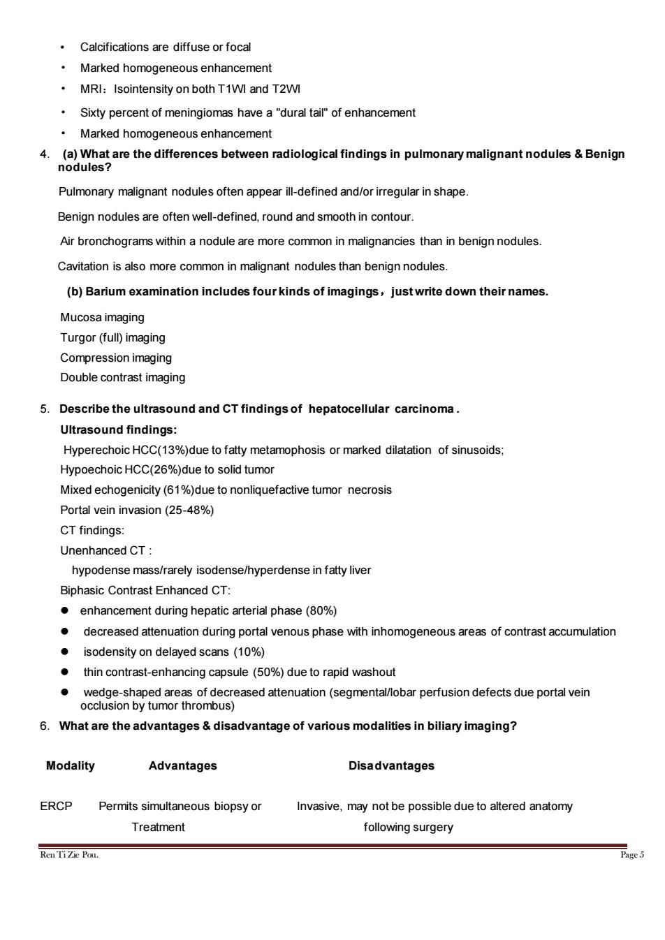
Calcifications are diffuse or focal Marked homogeneous enhancement MRI:Isointensity on both T1WI and T2WI Sixty percent of meningiomas have a"dural tail of enhancement Marked homogeneous enhancement 4.(a)What are the differences between radiological findings in pulmonary malignant nodules&Benign nodules? Pulmonary malignant nodules often appear ill-defined and/or irregular in shape. Benign nodules are often well-defined,round and smooth in contour. Air bronchograms within a nodule are more common in malignancies than in benign nodules Cavitation is also more common in malignant nodules than benign nodules (b)Barium examination includes four kinds of imagings,justwrite down their names Mucosa imaging Turgor(full)imaging Compression imaging Double contrast imaging 5.Describe the ultrasound and CT findings of hepatocellular carcinoma. Ultrasound findings: HyperechoicHCC(13%)due to fatty metamophosis or marked dilatation of sinusoids Hypoechoic HCC(26%)due to solid tumor Mixed echogenicity (6%due to nonliquefactive tumor necrosis Portal vein invasion (25-48%) CT findings: Unenhanced CT: hypodense mass/rarely isodense/hyperdense in fatty live Biphasic Contrast Enhanced CT: enhancement during hepatic arterial phase(80%) decreased attenuation during portal venous phase with inhomogeneous areas of contrast accumulation isodensity on delayed scans(10%) thin contrast-enhancing capsule(50%)due to rapid washout wedge-shaped areas of decreased attenuation(segmental/lobar perfusion defects due portal vein occlusion by tumor thrombus) 6.What are the advantages&disadvantage of various modalities in biliary imaging? Modality Advantages Disadvantages ERCP Permits simultaneous biopsy or Invasive.may not be possible due to altered anatomy Treatment following surgery RenTiZe Pou.Ren Ti Zie Pou. Page 5 • Calcifications are diffuse or focal • Marked homogeneous enhancement • MRI:Isointensity on both T1WI and T2WI • Sixty percent of meningiomas have a "dural tail" of enhancement • Marked homogeneous enhancement 4. (a) What are the differences between radiological findings in pulmonary malignant nodules & Benign nodules? Pulmonary malignant nodules often appear ill-defined and/or irregular in shape. Benign nodules are often well-defined, round and smooth in contour. Air bronchograms within a nodule are more common in malignancies than in benign nodules. Cavitation is also more common in malignant nodules than benign nodules. (b) Barium examination includes four kinds of imagings,just write down their names. Mucosa imaging Turgor (full) imaging Compression imaging Double contrast imaging 5. Describe the ultrasound and CT findings of hepatocellular carcinoma . Ultrasound findings: Hyperechoic HCC(13%)due to fatty metamophosis or marked dilatation of sinusoids; Hypoechoic HCC(26%)due to solid tumor Mixed echogenicity (61%)due to nonliquefactive tumor necrosis Portal vein invasion (25-48%) CT findings: Unenhanced CT : hypodense mass/rarely isodense/hyperdense in fatty liver Biphasic Contrast Enhanced CT: ⚫ enhancement during hepatic arterial phase (80%) ⚫ decreased attenuation during portal venous phase with inhomogeneous areas of contrast accumulation ⚫ isodensity on delayed scans (10%) ⚫ thin contrast-enhancing capsule (50%) due to rapid washout ⚫ wedge-shaped areas of decreased attenuation (segmental/lobar perfusion defects due portal vein occlusion by tumor thrombus) 6. What are the advantages & disadvantage of various modalities in biliary imaging? Modality Advantages Disadvantages ERCP Permits simultaneous biopsy or Invasive, may not be possible due to altered anatomy Treatment following surgery