正在加载图片...

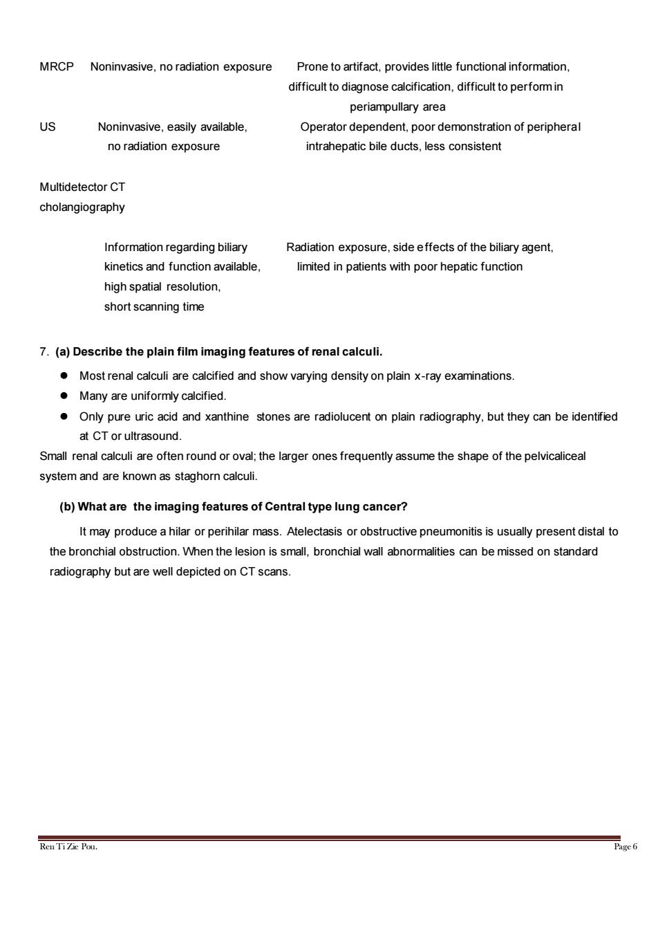
MRCP Noninvasive,no radiation exposure Prone to artifact,provides little functional information, difficult to diagnose calcification,difficult to perform in periampullary area US Noninvasive,easily available, Operator dependent,poor demonstration of peripheral no radiation exposure intrahepatic bile ducts,less consistent Multidetector CT cholangiography Information regarding biliary Radiation exposure,side effects of the biliary agent, kinetics and function available, limited in patients with poor hepatic function high spatial resolution, short scanning time 7.(a)Describe the plain film imaging features of renal calculi. Most renal calculi are calcified and show varying density on plain x-ray examinations. Many are uniformly calcified. Only pure uric acid and xanthine stones are radiolucent on plain radiography,but they can be identified at CT or ultrasound. Small renal calculi are often round or oval;the larger ones frequently assume the shape of the pelvicaliceal system and are known as staghorn calculi. (b)What are the imaging features of Central type lung cancer? It may produce a hilar or perihilar mass.Atelectasis or obstructive pneumonitis is usually present distal to the bronchial obstruction.When the lesion is small,bronchial wall abnormalities can be missed on standard radiography but are well depicted on CT scans. Ren TiZie Pou. Page 6Ren Ti Zie Pou. Page 6 MRCP Noninvasive, no radiation exposure Prone to artifact, provides little functional information, difficult to diagnose calcification, difficult to perform in periampullary area US Noninvasive, easily available, Operator dependent, poor demonstration of peripheral no radiation exposure intrahepatic bile ducts, less consistent Multidetector CT cholangiography Information regarding biliary Radiation exposure, side effects of the biliary agent, kinetics and function available, limited in patients with poor hepatic function high spatial resolution, short scanning time 7. (a) Describe the plain film imaging features of renal calculi. ⚫ Most renal calculi are calcified and show varying density on plain x-ray examinations. ⚫ Many are uniformly calcified. ⚫ Only pure uric acid and xanthine stones are radiolucent on plain radiography, but they can be identified at CT or ultrasound. Small renal calculi are often round or oval; the larger ones frequently assume the shape of the pelvicaliceal system and are known as staghorn calculi. (b) What are the imaging features of Central type lung cancer? It may produce a hilar or perihilar mass. Atelectasis or obstructive pneumonitis is usually present distal to the bronchial obstruction. When the lesion is small, bronchial wall abnormalities can be missed on standard radiography but are well depicted on CT scans