正在加载图片...
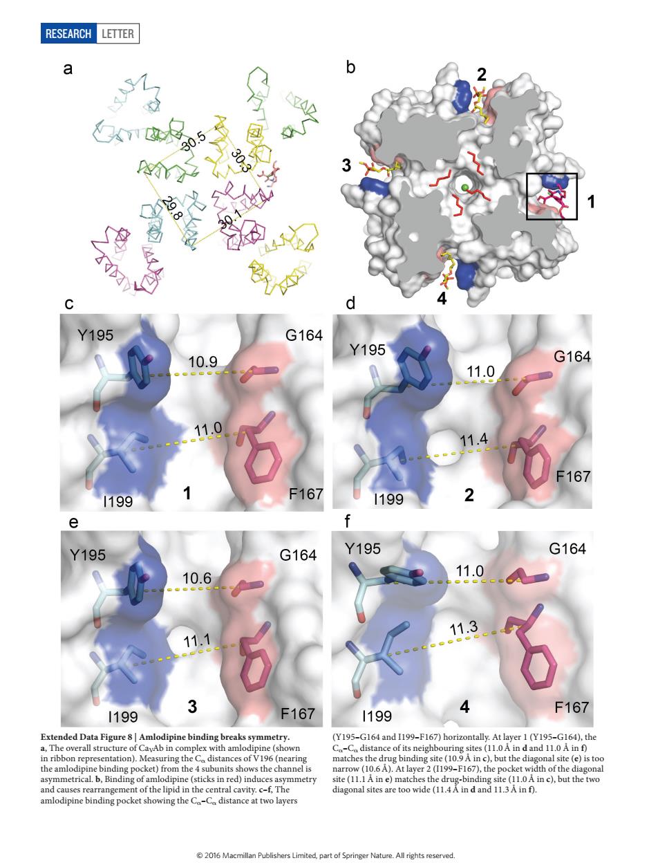
RESEARCH LETTER 7 学 /195 G164 195 10.9 G164 11.0 16 199 1199 1I G164 Y195 G164 10.6 11.0 11.3 F167 1199 F167 1199 nded Data Figure Aml dinine binding breaks s (Y195-G164 and I199-F167)horizontally At layer 1(Y195-G164),the nd and 11.0 A in ut t idth of the d metrical. e(n)ma n c),but the two mlodipine binding pocket showing the Co-Co distance at two layers 2016 Macmillan Publishers Limited part of Springer Nature.All tiehts reserved RESEARCH Letter Extended Data Figure 8 | Amlodipine binding breaks symmetry. a, The overall structure of CaVAb in complex with amlodipine (shown in ribbon representation). Measuring the Cα distances of V196 (nearing the amlodipine binding pocket) from the 4 subunits shows the channel is asymmetrical. b, Binding of amlodipine (sticks in red) induces asymmetry and causes rearrangement of the lipid in the central cavity. c–f, The amlodipine binding pocket showing the Cα–Cα distance at two layers (Y195–G164 and I199–F167) horizontally. At layer 1 (Y195–G164), the Cα–Cα distance of its neighbouring sites (11.0Å in d and 11.0 Å in f) matches the drug binding site (10.9Å in c), but the diagonal site (e) is too narrow (10.6Å). At layer 2 (I199–F167), the pocket width of the diagonal site (11.1Å in e) matches the drug-binding site (11.0Å in c), but the two diagonal sites are too wide (11.4Å in d and 11.3Å in f). © 2016 Macmillan Publishers Limited, part of Springer Nature. All rights reserved