正在加载图片...
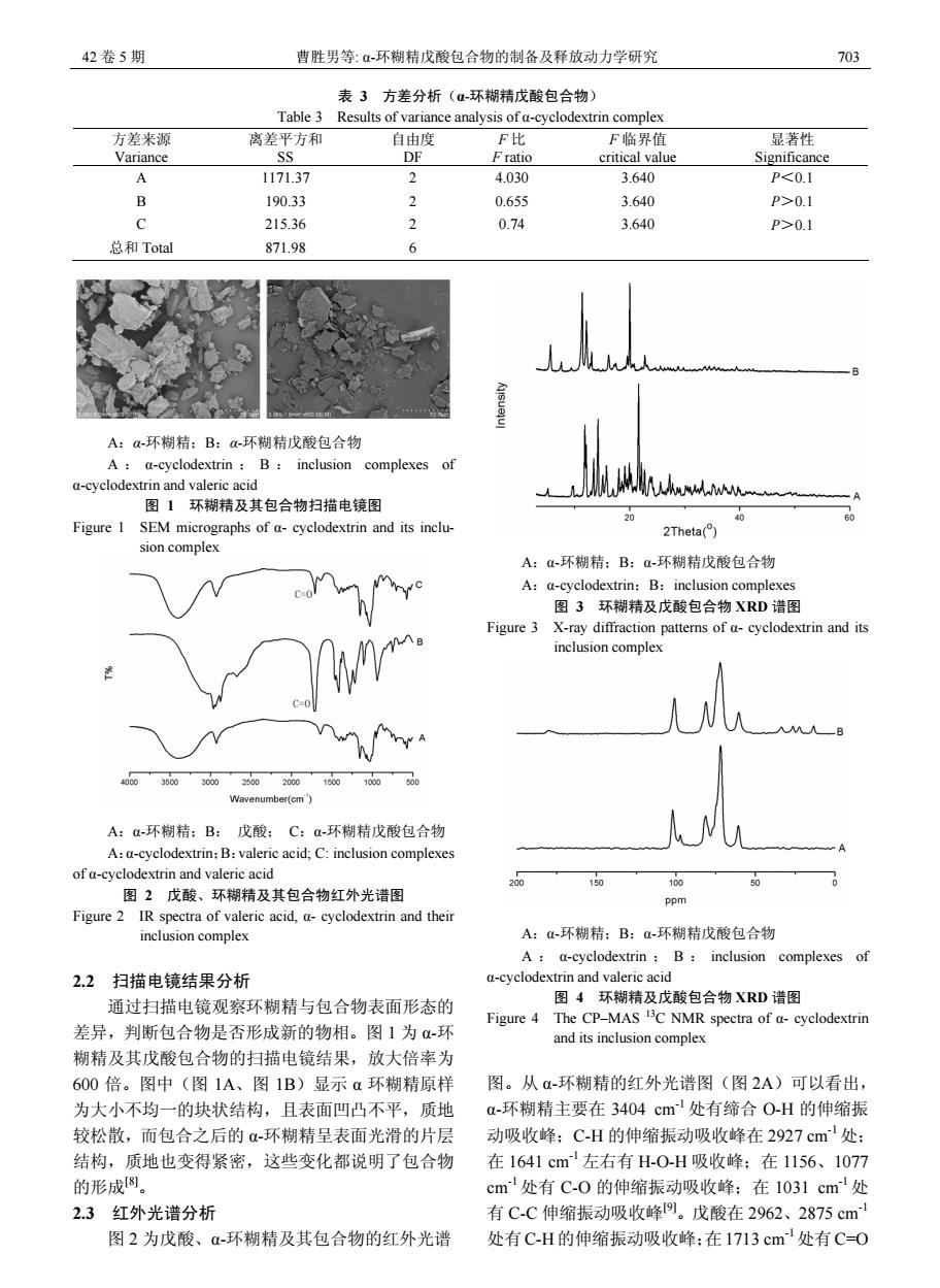
42卷5期 曹胜男等:α-环糊精戊酸包合物的制备及释放动力学研究 703 表3方差分析(α-环糊精戊酸包合物) Table 3 Results of variance analysis of a-cyclodextrin complex 方差来源 离差平方和 自由度 F比 F临界值 显著性 Variance SS DF Fratio critical value Significance A 1171.37 2 4.030 3.640 P<0.1 B 190.33 2 0.655 3.640 P>0.1 c 215.36 2 0.74 3.640 P>0.1 总和Total 871.98 6 A:a-环糊精:B:a-环糊精戊酸包合物 A a-cyclodextrin B:inclusion complexes of a-cyclodextrin and valeric acid 图1环糊精及其包合物扫描电镜图 20 Figure 1 SEM micrographs of a-cyclodextrin and its inclu- 2Theta() sion complex A:a-环糊精:B:a-环糊精戊酸包合物 A:a-cyclodextrin:B:inclusion complexes 图3环糊精及戊酸包合物XRD谱图 Figure 3 X-ray diffraction patterns of a-cyclodextrin and its inclusion complex 人N 20002300200 1800 100000 Wavenumber(cm' A:a-环糊精:B:戊酸:C:Q-环糊精戊酸包合物 A:a-cyclodextrin:B:valeric acid;C:inclusion complexes of a-cyclodextrin and valeric acid 300 460 100 图2戊酸、环糊精及其包合物红外光谱图 ppm Figure 2 IR spectra of valeric acid,a-cyclodextrin and their inclusion complex A:-环糊精:B:d-环糊精戊酸包合物 A a-cyclodextrin;B:inclusion complexes of 2.2扫描电镜结果分析 a-cyclodextrin and valeric acid 通过扫描电镜观察环糊精与包合物表面形态的 图4环糊精及戊酸包合物XRD谱图 Figure 4 The CP-MAS C NMR spectra of a-cyclodextrin 差异,判断包合物是否形成新的物相。图1为-环 and its inclusion complex 糊精及其戊酸包合物的扫描电镜结果,放大倍率为 600倍。图中(图1A、图1B)显示a环糊精原样 图。从a-环糊精的红外光谱图(图2A)可以看出, 为大小不均一的块状结构,且表面凹凸不平,质地 a-环糊精主要在3404cm'处有缔合O-H的伸缩振 较松散,而包合之后的α-环糊精呈表面光滑的片层 动吸收峰:C-H的伸缩振动吸收峰在2927cm'处: 结构,质地也变得紧密,这些变化都说明了包合物 在1641cm左右有H-0-H吸收峰:在1156、1077 的形成8。 cm处有C-0的伸缩振动吸收峰:在1031cm处 2.3红外光谱分析 有C-C伸缩振动吸收峰9。戊酸在2962、2875cm 图2为戊酸、α-环糊精及其包合物的红外光谱 处有C-H的伸缩振动吸收峰:在1713cm处有C=O42 卷 5 期 曹胜男等: α-环糊精戊酸包合物的制备及释放动力学研究 703 表 3 方差分析(α-环糊精戊酸包合物) Table 3 Results of variance analysis of α-cyclodextrin complex 方差来源 Variance 离差平方和 SS 自由度 DF F 比 F ratio F 临界值 critical value 显著性 Significance A 1171.37 2 4.030 3.640 P<0.1 B 190.33 2 0.655 3.640 P>0.1 C 215.36 2 0.74 3.640 P>0.1 总和 Total 871.98 6 A:α-环糊精;B:α-环糊精戊酸包合物 A : α-cyclodextrin ; B : inclusion complexes of α-cyclodextrin and valeric acid 图 1 环糊精及其包合物扫描电镜图 Figure 1 SEM micrographs of α- cyclodextrin and its inclusion complex A:α-环糊精;B: 戊酸; C:α-环糊精戊酸包合物 A:α-cyclodextrin;B:valeric acid; C: inclusion complexes of α-cyclodextrin and valeric acid 图 2 戊酸、环糊精及其包合物红外光谱图 Figure 2 IR spectra of valeric acid, α- cyclodextrin and their inclusion complex 2.2 扫描电镜结果分析 通过扫描电镜观察环糊精与包合物表面形态的 差异,判断包合物是否形成新的物相。图 1 为 α-环 糊精及其戊酸包合物的扫描电镜结果,放大倍率为 600 倍。图中(图 1A、图 1B)显示 α 环糊精原样 为大小不均一的块状结构,且表面凹凸不平,质地 较松散,而包合之后的 α-环糊精呈表面光滑的片层 结构,质地也变得紧密,这些变化都说明了包合物 的形成[8]。 2.3 红外光谱分析 图 2 为戊酸、α-环糊精及其包合物的红外光谱 A:α-环糊精;B:α-环糊精戊酸包合物 A:α-cyclodextrin;B:inclusion complexes 图 3 环糊精及戊酸包合物 XRD 谱图 Figure 3 X-ray diffraction patterns of α- cyclodextrin and its inclusion complex A:α-环糊精;B:α-环糊精戊酸包合物 A : α-cyclodextrin ; B : inclusion complexes of α-cyclodextrin and valeric acid 图 4 环糊精及戊酸包合物 XRD 谱图 Figure 4 The CP–MAS 13C NMR spectra of α- cyclodextrin and its inclusion complex 图。从 α-环糊精的红外光谱图(图 2A)可以看出, α-环糊精主要在 3404 cm-1 处有缔合 O-H 的伸缩振 动吸收峰;C-H 的伸缩振动吸收峰在 2927 cm-1 处; 在 1641 cm-1 左右有 H-O-H 吸收峰;在 1156、1077 cm -1 处有 C-O 的伸缩振动吸收峰;在 1031 cm-1处 有 C-C 伸缩振动吸收峰[9]。戊酸在 2962、2875 cm-1 处有 C-H 的伸缩振动吸收峰;在 1713 cm-1处有 C=O