Stain Protocols-BIOL 2420 Simple Stain Simple stains provide a quick and easy way to determine cell shape,size,and arrangement. 1.Perform a bacterial smear,as discussed in Figure 3-52 on page 150 of your lab manual. 2.Saturate the smear with basic dye for approximately 1 minute.You may use crystal violet safranin,or methylene blue. 3.Rinse the slide gently with water. 4.Carefully blot dry with bibulous paper 5.Observe the slide under the microscope,using proper microscope technique. Negative Stain anism'scelular morphology.Since the in any way. mMis the uns ed c 1.Place a single drop of nigrosin on a clean microscope slide,adjacent to the frosted edge 2.Usinga flamed loop and steri i remove some organism from yo and mix it into the dr rop of nigrosin. Be ue there are no large clumps of organism,but try to avoid spreading the drop. 3.Place the end of another clean microscope slide at an angle to the end of the slide containing the organism and spread the drop out into a film.This is done by contacting the drop of nigro slide and using the capillary action of the s the s 4.Allow the film to air dry. 5.Observe the slide under the microscope,using proper microscope technique. Smith-2010 Page1of4
Stain Protocols – BIOL 2420 Smith – 2010 Page 1 of 4 Simple Stain Simple stains provide a quick and easy way to determine cell shape, size, and arrangement. 1. Perform a bacterial smear, as discussed in Figure 3-52 on page 150 of your lab manual. 2. Saturate the smear with basic dye for approximately 1 minute. You may use crystal violet, safranin, or methylene blue. 3. Rinse the slide gently with water. 4. Carefully blot dry with bibulous paper. 5. Observe the slide under the microscope, using proper microscope technique. Negative Stain Negative staining is an excellent way to determine an organism’s cellular morphology. Since the cells themselves are not stained, their morphology is not distorted in any way. The nigrosin provides a dark background against which the shapes of the unstained cells are clearly visible. This method provides a high degree of contrast not available in most other staining procedures. 1. Place a single drop of nigrosin on a clean microscope slide, adjacent to the frosted edge. 2. Using a flamed loop and sterile technique, remove some organism from your tube or plate and mix it into the drop of nigrosin. Be sure there are no large clumps of organism, but try to avoid spreading the drop. 3. Place the end of another clean microscope slide at an angle to the end of the slide containing the organism and spread the drop out into a film. This is done by contacting the drop of nigrosin with the clean microscope slide and using the capillary action of the dye/microscope slide to spread the nigrosin across the smear: 4. Allow the film to air dry. 5. Observe the slide under the microscope, using proper microscope technique

Stain Protocols-BIOL 2420 Gram Stain 1.Perform a bacterial smear,as discussed in Figure 3-52 on page 150 of your lab manual. 2.Saturate the smear with crystal violet for 1 minute 3.Rinse the slide gently with water. 4.Saturate the smear with iodine for I minute. 5.Rinse the slide gently with water. 6.Decolorize with Gram decolorizer(acetone/alcohol)for 3-5 seconds ONLY;if you leave the decolorizer on too long,it will bleach the crystal violet out of a gram positive cell! 7.Rinse the slide gently with water. 8.Counterstain with safranin for 1 minute 9.Rinse the slide gently with water 10.Carefully blot the slide dry with bibulous paper. 11.Observe the slide under the microscope,using proper microscope technique. With proper staining technique.. Gram positive bacteria will stain purple. Gram negative bacteria will stain red/pink TIP:When making a smear of an unknown organism to Gram stain,place three nmon an pole yor staining technique,it will be reflected in the known organisms,and you'll know you need to do the stain again to get accurate esults. Smith-2010 Page2of4
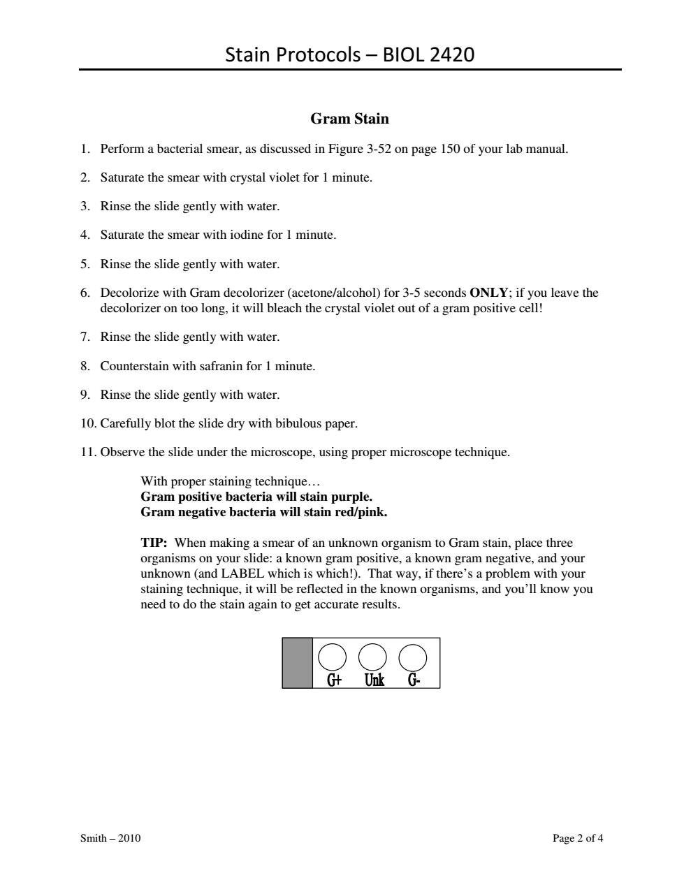
Stain Protocols – BIOL 2420 Smith – 2010 Page 2 of 4 Gram Stain 1. Perform a bacterial smear, as discussed in Figure 3-52 on page 150 of your lab manual. 2. Saturate the smear with crystal violet for 1 minute. 3. Rinse the slide gently with water. 4. Saturate the smear with iodine for 1 minute. 5. Rinse the slide gently with water. 6. Decolorize with Gram decolorizer (acetone/alcohol) for 3-5 seconds ONLY; if you leave the decolorizer on too long, it will bleach the crystal violet out of a gram positive cell! 7. Rinse the slide gently with water. 8. Counterstain with safranin for 1 minute. 9. Rinse the slide gently with water. 10. Carefully blot the slide dry with bibulous paper. 11. Observe the slide under the microscope, using proper microscope technique. With proper staining technique… Gram positive bacteria will stain purple. Gram negative bacteria will stain red/pink. TIP: When making a smear of an unknown organism to Gram stain, place three organisms on your slide: a known gram positive, a known gram negative, and your unknown (and LABEL which is which!). That way, if there’s a problem with your staining technique, it will be reflected in the known organisms, and you’ll know you need to do the stain again to get accurate results

Stain Protocols-BIOL 2420 Capsule Stain 1.Place a single drop of India ink on a clean microscope slide,adjacent to the frosted adge 2.Using a flamed loop and sterile technique,remove some K.pneumoniae (or the organism you want to stain)from your tube or plate and mix it into the drop of India ink.Be sure there are no large clumps of organism,but try to avoid spreading the drop. 3.Place the end of another clean microscope slide at an angle to the end of the slide containing the organism.Spread out the drop out into a film.This is done by contacting the drop of India ink with the clean microscope slide and using the capillary action of the dye/slide to spread the India ink across the smear.Refer to the Negative Stain portion of this handout for a diagram. 4.Allow the film to air dry.DO NOT heat or blot dry!!!!Heat will melt the capsule! 5.Saturate the slide with crystal violet for 1 minute. 6.Rinse the slide gently with water. 7.Allow the slide to air dry.DO NOT heat or blot dry!!!!Heat will melt the capsule! 8.Observe the slide under the microscope,using proper microscope technique The background will be dark. The bacterial cells will be stained purple. The capsule(if present)will appear clear against the dark background. background capsule bacterium 0 TIP:If you are stainin ism he sure ur culture is several days old.Young fresh cult on't ha veloped capsules yet. Smith-2010 Page3of4
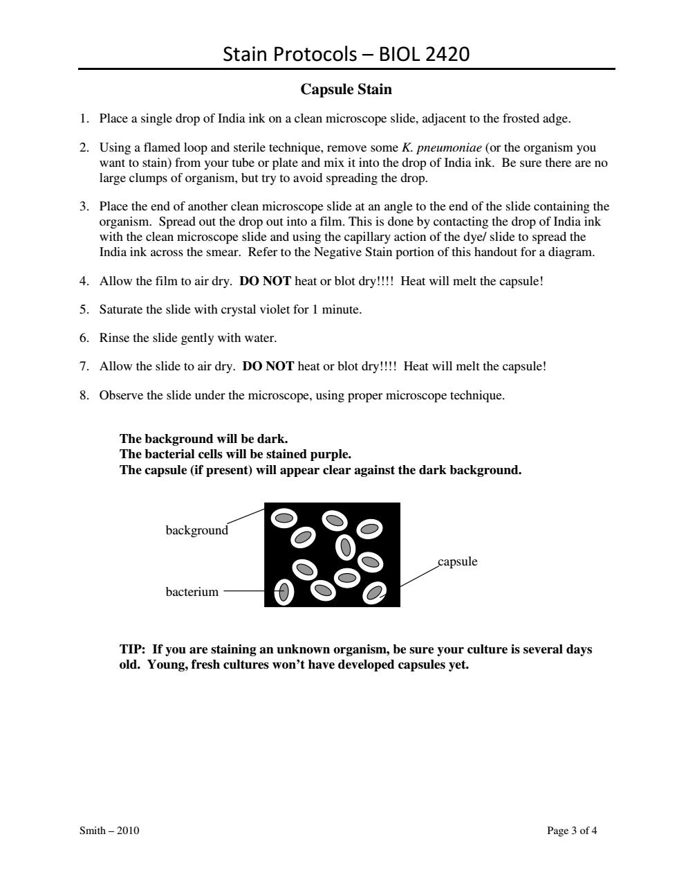
Stain Protocols – BIOL 2420 Smith – 2010 Page 3 of 4 Capsule Stain 1. Place a single drop of India ink on a clean microscope slide, adjacent to the frosted adge. 2. Using a flamed loop and sterile technique, remove some K. pneumoniae (or the organism you want to stain) from your tube or plate and mix it into the drop of India ink. Be sure there are no large clumps of organism, but try to avoid spreading the drop. 3. Place the end of another clean microscope slide at an angle to the end of the slide containing the organism. Spread out the drop out into a film. This is done by contacting the drop of India ink with the clean microscope slide and using the capillary action of the dye/ slide to spread the India ink across the smear. Refer to the Negative Stain portion of this handout for a diagram. 4. Allow the film to air dry. DO NOT heat or blot dry!!!! Heat will melt the capsule! 5. Saturate the slide with crystal violet for 1 minute. 6. Rinse the slide gently with water. 7. Allow the slide to air dry. DO NOT heat or blot dry!!!! Heat will melt the capsule! 8. Observe the slide under the microscope, using proper microscope technique. The background will be dark. The bacterial cells will be stained purple. The capsule (if present) will appear clear against the dark background. background capsule bacterium TIP: If you are staining an unknown organism, be sure your culture is several days old. Young, fresh cultures won’t have developed capsules yet

Stain Protocols-BIOL 2420 Acid-Fast Stain 1.Perform a bacterial smear,as discussed in Figure 3-52 on page 150 of your lab manual. 2.Place a small piece of bibulous paper over the smear and saturate the paper with carbolfuchsin. 3.Heat the slide gently over the Bunsen burner for 5 minutes.Be sure to keep the bibulous paper saturated with carbolfuchsin during heating.If the slide is steaming,you're okay;if it stops steaming,add more carbolfuchsin! 4.Remove the bibulous paper from the slide,and rinse the slide gently with water.Dispose of the used bibulous paper in the trash.DO NOT leave the bibulous paper in the sink or drain! 5.Decolorize the slide with acid-alcohol until the rinsate runs clear. 6.Rinse the slide gently with water. 7.Counterstain with methylene blue for 2 minutes. 8.Rinse the slide gently with water. 9.Carefully blot the slide dry with bibulous paper. 10.Observe the slide under the microscope,using proper microscope technique. Acid-fast cells will stain fuchsia.Non-acid-fast cells will stain blue. Endospore Stain 1.Perform a bacterial smear of Bacillus or the organism you want to stain,as discussed in Figure 3-52 on page 150 of your lab manual. 2.Place a small piece of bibulous paper over the smear.Saturate the paper with malachite green. 3.Heat the slide gently over the Bunsen burner for 5 minutes.Be sure to keep the bibulous paper saturated with malachite green during heating.If the slide is steaming,you're okay;if it stops steaming,add more malachite green! 4.Remove the bibulous paper from the slide,and rinse the slide gently with water.Dispose of the used bibulous paper in the trash.DO NOT leave the bibulous paper in the sink or drain! Parent cell or 5.Counterstain with safranin for 2 minutes. bacterium(red) 6.Rinse the slide gently with water. 7.Carefully blot the slide dry with bibulous paper. Endospore (green) 8.Observe the slide under the microscope,using proper microscope technique. Endospores will stain green.Parent cells will stain red. Smith-2010 Page 4 of 4
Stain Protocols – BIOL 2420 Smith – 2010 Page 4 of 4 Acid-Fast Stain 1. Perform a bacterial smear, as discussed in Figure 3-52 on page 150 of your lab manual. 2. Place a small piece of bibulous paper over the smear and saturate the paper with carbolfuchsin. 3. Heat the slide gently over the Bunsen burner for 5 minutes. Be sure to keep the bibulous paper saturated with carbolfuchsin during heating. If the slide is steaming, you’re okay; if it stops steaming, add more carbolfuchsin! 4. Remove the bibulous paper from the slide, and rinse the slide gently with water. Dispose of the used bibulous paper in the trash. DO NOT leave the bibulous paper in the sink or drain! 5. Decolorize the slide with acid-alcohol until the rinsate runs clear. 6. Rinse the slide gently with water. 7. Counterstain with methylene blue for 2 minutes. 8. Rinse the slide gently with water. 9. Carefully blot the slide dry with bibulous paper. 10. Observe the slide under the microscope, using proper microscope technique. Acid-fast cells will stain fuchsia. Non-acid-fast cells will stain blue. Endospore Stain 1. Perform a bacterial smear of Bacillus or the organism you want to stain, as discussed in Figure 3-52 on page 150 of your lab manual. 2. Place a small piece of bibulous paper over the smear. Saturate the paper with malachite green. 3. Heat the slide gently over the Bunsen burner for 5 minutes. Be sure to keep the bibulous paper saturated with malachite green during heating. If the slide is steaming, you’re okay; if it stops steaming, add more malachite green! 4. Remove the bibulous paper from the slide, and rinse the slide gently with water. Dispose of the used bibulous paper in the trash. DO NOT leave the bibulous paper in the sink or drain! 5. Counterstain with safranin for 2 minutes. 6. Rinse the slide gently with water. 7. Carefully blot the slide dry with bibulous paper. 8. Observe the slide under the microscope, using proper microscope technique. Endospores will stain green. Parent cells will stain red. Parent cell or bacterium (red) Endospore (green)