Respiratory infections and interstitial disease 2008-12
Respiratory infections and interstitial disease 2008-12

Pulmonary veins Bronchiole Visceral pleura Interlobular septum Pulmonary artery Lymphatics Fig.1.-Line drawing ot a secondary pulmorary lobule.Borders of lobule are interlobular septa.At center of each lcbule isa bronchiole and a pulmonary artery (b).Pulmonary vein (red)run in interlobular septa.Lymphatics (green are found in interlobular septa and in central or axial interstitium that surrounds bronchovascular bundles
Distribution ·Lobar pneumonia ·Bronchopneumonia Extensive bilateral pneumonia Bilateral lower lobe pneumonia Peripheral pneumonia
Distribution • Lobar pneumonia • Bronchopneumonia • Extensive bilateral pneumonia • Bilateral lower lobe pneumonia • Peripheral pneumonia
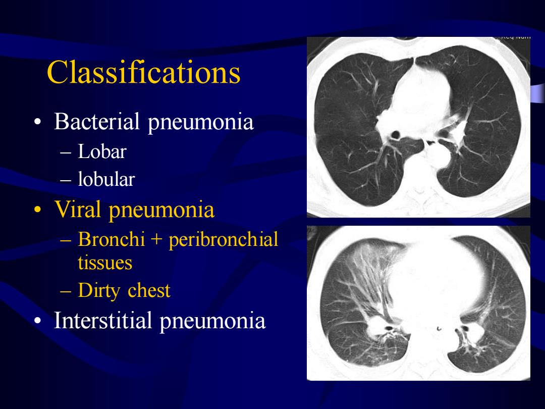
Classifications Bacterial pneumonia Lobar lobular ·Viral pneumonia Bronchi peribronchial tissues Dirty chest Interstitial pneumonia
Classifications • Bacterial pneumonia – Lobar – lobular • Viral pneumonia – Bronchi + peribronchial tissues – Dirty chest • Interstitial pneumonia
3910 ast
器 70 r:ned 122 Ser Redm 12
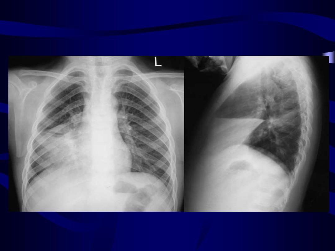
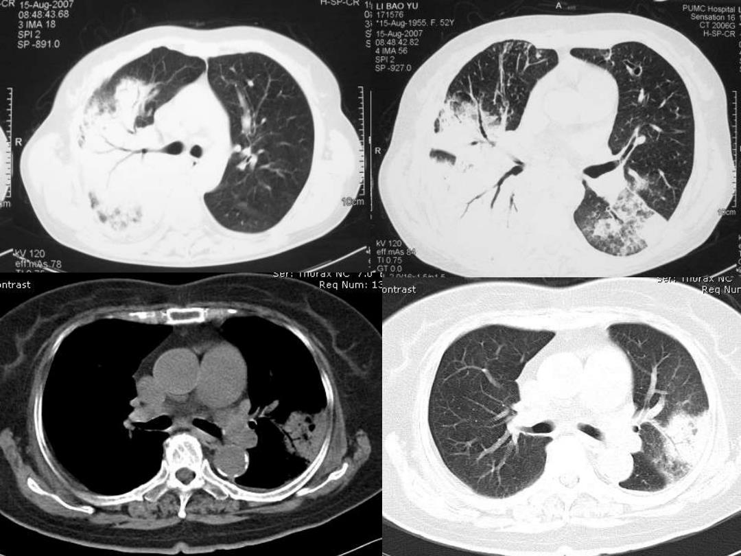
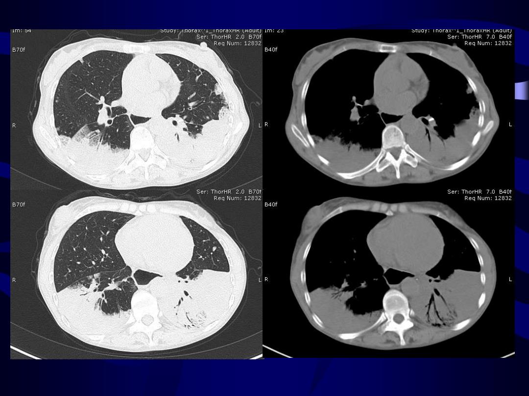
Lobar pneumonia ·Consolidation ·Airway sign ·Lack of volume loss
Lobar pneumonia • Consolidation • Airway sign • Lack of volume loss

Complications ·Abscess:脓肿-见后 ·Cavitary necrosis:空洞样坏死 ·Pneumothorax:气胸 ·Pyopneumothorax:脓气胸 。Bronchopleural fistula:支气管胸膜瘘
Complications • Abscess:脓肿-见后… • Cavitary necrosis:空洞样坏死 • Pneumothorax:气胸 • Pyopneumothorax:脓气胸 • Bronchopleural fistula:支气管胸膜瘘

是 部儒