
第八章 核糖体
第八章 核糖体
核糖体(核糖核蛋白体) 。没有膜包被的细胞器 。几乎存在于一切细胞中(哺乳动物成熟的红细胞 等极个别高度分化的细胞除外)
核糖体(核糖核蛋白体) ⚫ 没有膜包被的细胞器 ⚫ 几乎存在于一切细胞中(哺乳动物成熟的红细胞 等极个别高度分化的细胞除外)
核糖体的类型与化学组成 形态结构 颗粒状 ·存在方式 ■游离、附着 ·化学组成 ■r蛋白质:约占13 ■rRNA:约占2/3
一、核糖体的类型与化学组成 ⚫ 形态结构 ◼ 颗粒状 ⚫ 存在方式 ◼ 游离、附着 ⚫ 化学组成 ◼ r蛋白质:约占1/3 ◼ rRNA:约占2/3
·类型 原核生物的核糖体:70S(30S,50S) ■真核生物的核糖体:80S(40S,60S) 。功能:蛋白质的合成场所 ·数量、分布与蛋白质合成程度有关
⚫ 类型 ◼ 原核生物的核糖体:70S(30S,50S) ◼ 真核生物的核糖体:80S(40S,60S) ⚫ 功能:蛋白质的合成场所 ⚫ 数量、分布与蛋白质合成程度有关
二、蛋白质的合成 ·多聚核糖体 ●蛋白质的合成 ■肽链的起始 ■肽链的延伸 ■肽链的终止
二、蛋白质的合成 ⚫ 多聚核糖体 ⚫ 蛋白质的合成 ◼ 肽链的起始 ◼ 肽链的延伸 ◼ 肽链的终止
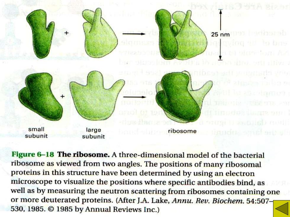
25 nm small large ribosome subunit subunit Figure 6-18 The ribosome.A three-dimensional model of the bacterial ribosome as viewed from two angles.The positions of many ribosomal proteins in this structure have been determined by using an electron microscope to visualize the positions where specific antibodies bind,as well as by measuring the neutron scattering from ribosomes containing one or more deuterated proteins.(After J.A.Lake,Annu.Rev.Biochem.54:507- 530,1985.1985 by Annual Reviews Inc.)

70s 80S MW2.500.000 MW4,200,000 509 30S 40S MW1.600.000 MW900.000 MW2.800.000 MW1,400.000 5S rRNA 23S rRNA 16S rRNA 5S rRNA 28S rRNA 5.8S rRNA 18S rRNA 120 120 160 nucleotides 2900 1540 nucleotides nucleotides 1900 nucleotides nucleotides nucleotides 4700 nucleotides 34 proteins 21 proteins -49 proteins ~33 proteins PROCARYOTIC RIBOSOME EUCARYOTIC RIBOSOME Figure 6-20 A comparison of the structures of procaryotic and eucaryotic ribosomes.Ribosomal

Figure 7-26 RNA-binding sites in the ribosome.Each ribosome has a binding site for mRNA and three binding sites for tRNA,the A-,P-,and E-sites(short for E-site P-site A-site aminoacyl-tRNA,peptidyl-tRNA,and exit, respectively).(A)This representation ofa ribosome,which will be used in subse- large ribosomal quent figures,is highly schematic.(B)A subunit model of the procaryote ribosome that depicts the arrangement of the mRNA small ribosoma (orange beads)and the positions of one subunit tRNA molecule in the A-site of the ribo- some (pink),one tRNA molecule in the P-site of the ribosome (green),and one tRNA molecule in the E-site of the ribo- some(yellow).In this model the large ribosomal subunit is light blue,and the small subunit is light green.Although all B three tRNA sites are shown occupied here, during the process of protein synthesis not more than two of these sites contain tRNA molecules at any one time (see Figure 7-27).(B,courtesy of Joachim Frank, Yanhong Li,and Rajer