☒无法显示该图别 五 肺肿瘤 lung tumor 原发性 良性 恶性原发性支气管肺癌 转移性 重庆医科大学格床医学二系、医学彩德学 The department of second clinical medicine&Medical imaging of CoUMs
五 肺 肿 瘤 lung tumor • 原发性 良性 恶性 原发性支气管肺癌 • 转移性
(一)原发性支气管肺癌(primary bronchogenic carcinoma) [临床与病理 组织学类型 病理分型 小细胞癌(small cell lung cancer)) ● 中心型肺癌 非小细胞肺癌(non-small cell lung cancer) (squamous carcinoma) 外围型肺癌 腺癌(adenocarcinoma 细支气管肺泡癌 腺鳞癌(adenosquamous carcinoma) 大细胞癌(large cell carcinoma 重庆医科大学格床医学二系、医学影德学自 The department of socond clInical medicine Medical Imoging of COLMs
(一)原发性支气管肺癌(primary bronchogenic carcinoma) [临床与病理] 组织学类型 鳞癌(squamous carcinoma) 腺癌(adenocarcinoma) 腺鳞癌(adenosquamous carcinoma) 大细胞癌(large cell carcinoma) 病理分型 • 中心型肺癌 • 外围型肺癌 • 细支气管肺泡癌 小细胞癌(small cell lung cancer) 非小细胞肺癌(non-small cell lung cancer)
primary bronchogenic carcinoma 生长方式: 中心型 管内型 癌瘤向管腔内生长,阻塞支气管 管壁型 癌瘤沿支气管壁浸润生长,引起支气管壁增厚狭窄甚至梗 阻 管外型 癌瘤穿透支气管壁向外生长,形成肿块 外围型 侵入肺内形成肿块 细支气管肺泡癌 沿肺泡壁生长,经支气管及淋巴管播散 重庆送头学福床医 一退像学 The department of second clinical medicine Medical imaging of CouMs
生长方式: • 中心型 管内型 癌瘤向管腔内生长,阻塞支气管 管壁型 癌瘤沿支气管壁浸润生长,引起支气管壁增厚狭窄甚至梗 阻 管外型 癌瘤穿透支气管壁向外生长,形成肿块 • 外围型 侵入肺内形成肿块 • 细支气管肺泡癌 沿肺泡壁生长,经支气管及淋巴管播散 primary bronchogenic carcinoma

primary bronchogenic carcinoma 临床表现 。早期,阴性 ·上腔静脉阻塞综合征 。呼吸道症状:咳 喉返神经及膈神经麻痹 嗽、咯血、胸痛、 咳痰、呼吸困难等 重庆医科大学格床医学二系、医学影德学自 The department of second clinleal medicine Medical Imoging of CoUMs
primary bronchogenic carcinoma 临床表现 • 早期,阴性 • 呼吸道症状:咳 嗽、咯血、胸痛、 咳痰、呼吸困难等 • 上腔静脉阻塞综合征 • 喉返神经及膈神经麻痹
primary bronchogenic carcinoma [影像学表现] 。X线表现 CT表现 。] MRI表现 重庆医科大学格床医学二系、医学彩德学导 The department of second clinical medicine&Medical imaging of CoUMs
[影像学表现] • X线表现 • CT表现 • MRI表现 primary bronchogenic carcinoma
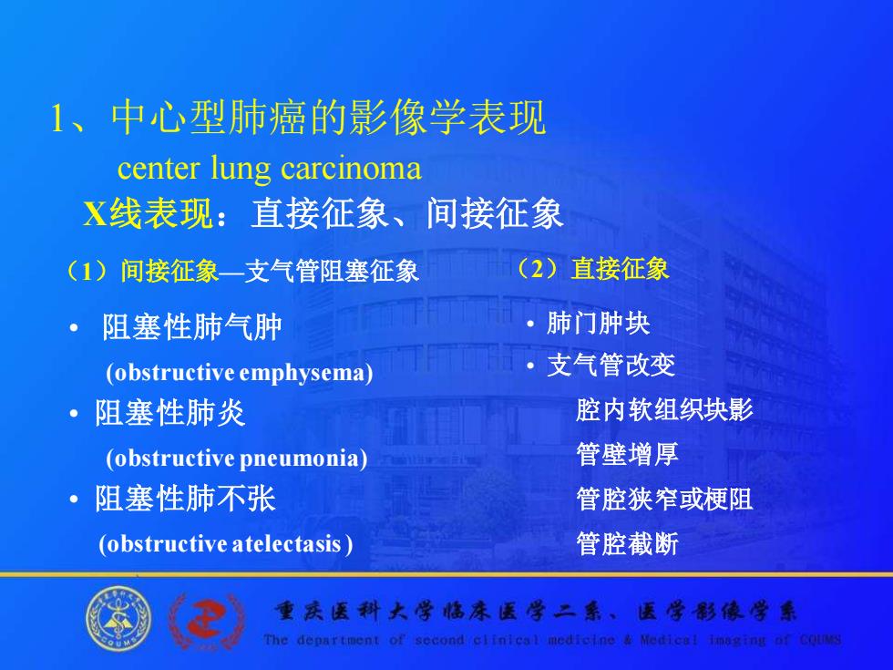
1、中心型肺癌的影像学表现 center lung carcinoma X线表现:直接征象、间接征象 (1)间接征象一支气管阻塞征象 (2)直接征象 阻塞性肺气肿 ·肺门肿块 (obstructive emphysema) ·支气管改变 阻塞性肺炎 腔内软组织块影 (obstructive pneumonia) 管壁增厚 ·阻塞性肺不张 管腔狭窄或梗阻 (obstructive atelectasis 管腔截断 重庆医科大学倦床医学二系、医学形像学 The department of second clinleal medicine Medical Imoging of CoUMs
1、中心型肺癌的影像学表现 center lung carcinoma X线表现:直接征象、间接征象 (1)间接征象—支气管阻塞征象 • 阻塞性肺气肿 (obstructive emphysema) • 阻塞性肺炎 (obstructive pneumonia) • 阻塞性肺不张 (obstructive atelectasis ) (2)直接征象 • 肺门肿块 • 支气管改变 腔内软组织块影 管壁增厚 管腔狭窄或梗阻 管腔截断
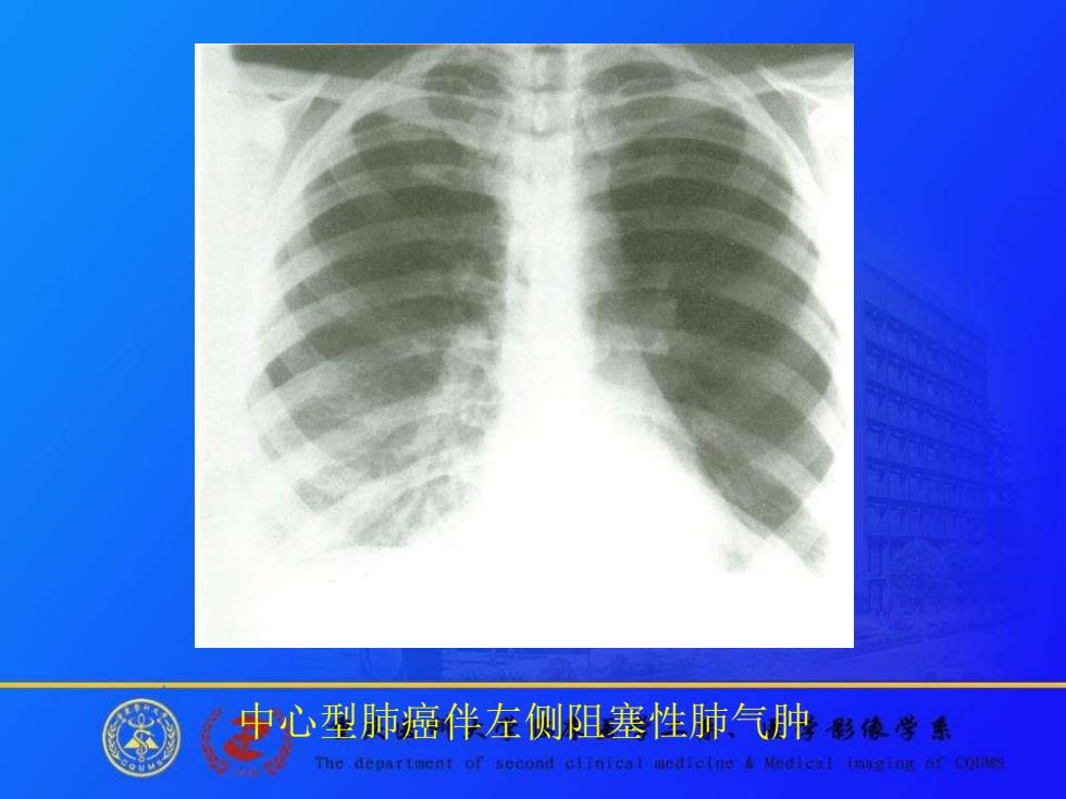
中心型肺癌伴左侧阻塞性肺气肿移像学◆ The department of second clinical medicine Medical imaging of CoUMs
中心型肺癌伴左侧阻塞性肺气肿
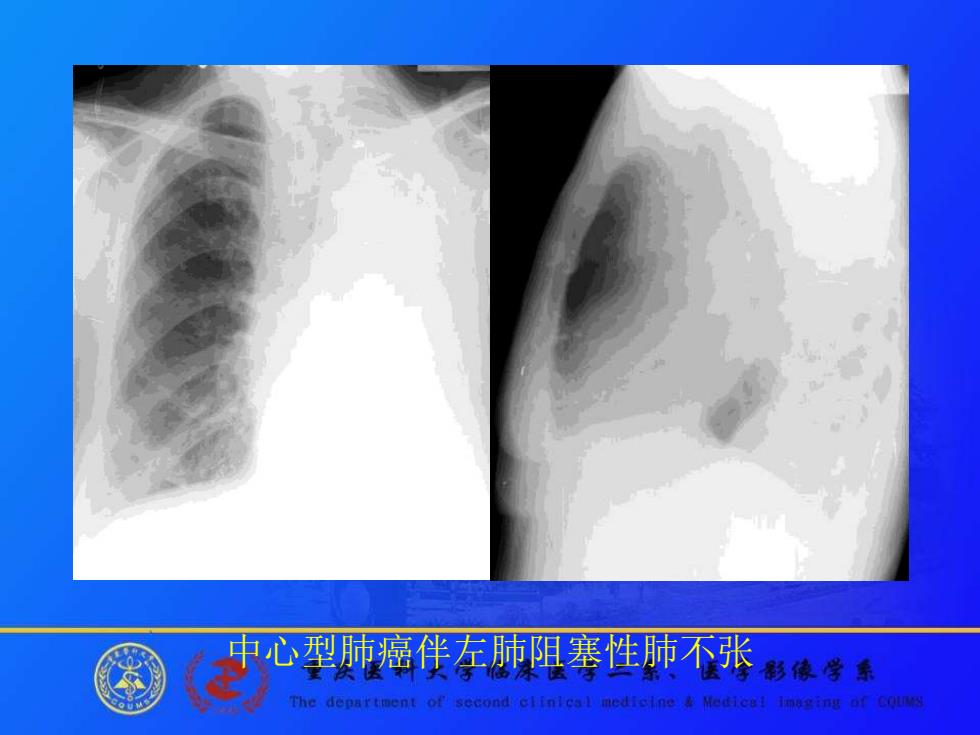
中心型肺癌伴左肺阻塞性肺不张 影像学乐 The department of second clinleal medicine Medical Imoging of CoUMs
中心型肺癌伴左肺阻塞性肺不张
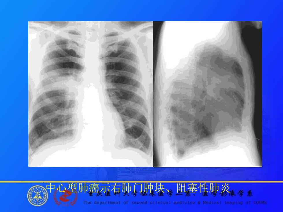
中心型肺癌示右肺门肿块、阻塞性肺炎学 The department of second clinical medicine Medical imaging of CoUMs
中心型肺癌示右肺门肿块、阻塞性肺炎
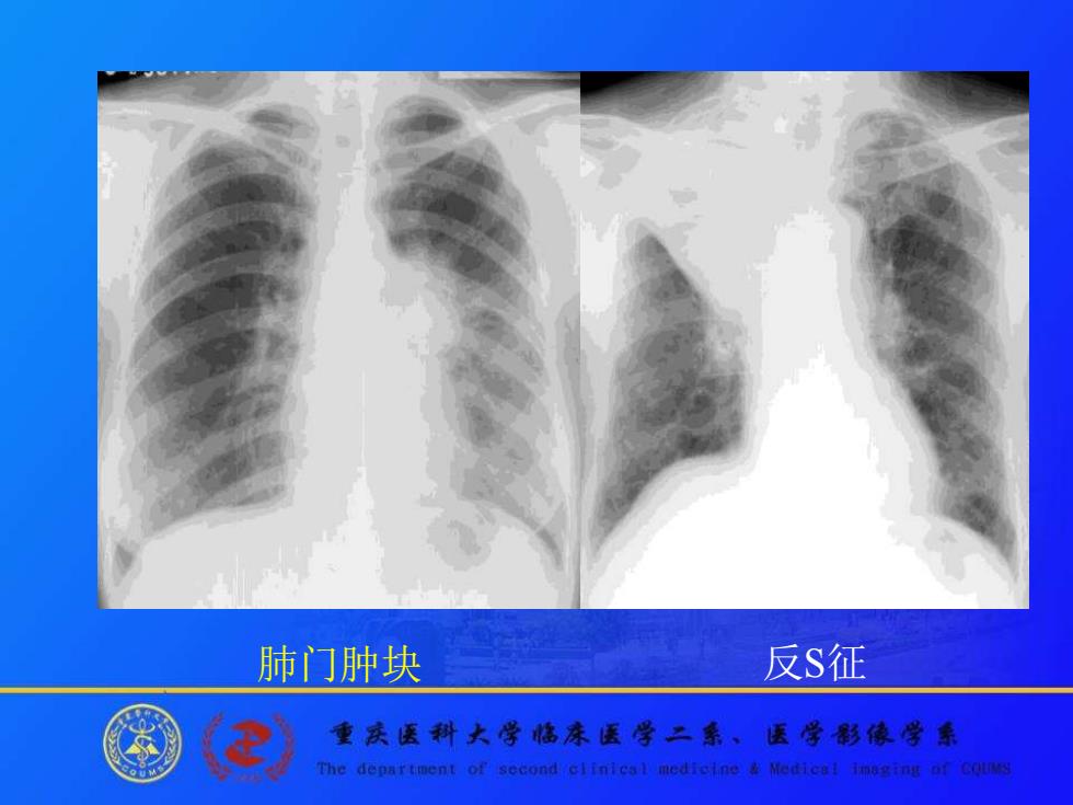
肺门肿块 反S征 ® 重庆医科大学格床医学二系、医学影德学乐 The department of second clinieal mediclne Medical Imoging of CoUMs
肺门肿块 反S征